

STAT5A Primary Antibody
Item Information
Catalog #
Size
Price
Description
The protein encoded by this gene is a member of the STAT family of transcription factors. In response to cytokines and growth factors, STAT family members are phosphorylated by the receptor associated kinases, and then form homo- or heterodimers that translocate to the cell nucleus where they act as transcription activators. This protein is activated by, and mediates the responses of many cell ligands, such as IL2, IL3, IL7 GM-CSF, erythropoietin, thrombopoietin, and different growth hormones. Activation of this protein in myeloma and lymphoma associated with a TEL/JAK2 gene fusion is independent of cell stimulus and has been shown to be essential for the tumorigenesis. The mouse counterpart of this gene is found to induce the expression of BCL2L1/BCL-X(L), which suggests the antiapoptotic function of this gene in cells.
Product Overview
Entrez GenelD
6776
Aliases
MGF; STAT5
Clone#
6D4
Host / Isotype
Mouse / IgG1
Species Reactivity
Human
Immunogen
Purified recombinant fragment of human STAT5A (AA: 583-794 ) expressed in E. Coli.
Formulation
Purified antibody in PBS with 0.05% sodium azide
Storage
Store at 4°C short term. Aliquot and store at -20°C long term. Avoid freeze/thaw cycles.
Product Applications
WB (Western Blot)
1/500 - 1/2000
IHC_P(Immunohistochemistry)
1/200 - 1/1000
ICC (Immunocytochemistry)
1/50
FCM (Flow Cytometry)
1/200 - 1/400
ELISA
Propose dilution 1/10000
References
1.Cancer Res. 2011 May 15;71(10):3720-31.
2.Blood. 2011 Mar 24;117(12):3409-20.
2.Blood. 2011 Mar 24;117(12):3409-20.
Product Image
Western Blot

Figure 1: Western blot analysis using STAT5A mAb against human STAT5A recombinant protein. (Expected MW is 49.3 kDa)
Western Blot
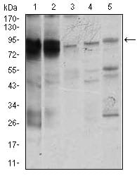
Figure 2: Western blot analysis using STAT5A mouse mAb against K562 (1), MOLT4 (2), HeLa (3), Jurkat (4), and A431 (5) cell lysate.
Immunofluorescence analysis
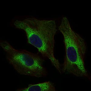
Figure 3: Immunofluorescence analysis of Hela cells using STAT5A mouse mAb (green). Blue: DRAQ5 fluorescent DNA dye.
Flow cytometric

Figure 4: Flow cytometric analysis of K562 cells using STAT5A mouse mAb (green) and negative control (red).
Immunohistochemical analysis

Figure 5: Immunohistochemical analysis of paraffin-embedded ovarian cancer tissues using STAT5A mouse mAb with DAB staining.
Immunohistochemical analysis

Figure 6: Immunohistochemical analysis of paraffin-embedded rectum cancer tissues using STAT5A mouse mAb with DAB staining.
Elisa

Black line: Control Antigen (100 ng); Purple line: Antigen(10ng); Blue line: Antigen (50 ng); Red line: Antigen (100 ng);
For Research Use Only. Not for use in diagnostic procedures.