

STAT3 Primary Antibody
Item Information
Catalog #
Size
Price
Description
The Stat3 transcription factor is an important signaling molecule for many cytokines and growth-factor receptors and is required for murine fetal development . Stat3 is constitutively activated in a number of human tumors and possesses oncogenic potential and anti-apoptotic activities. Stat3 is activated by phosphorylation at Tyr705, which induces dimerization, nuclear translocation and DNA binding. Transcriptional activation seems to be regulated by phosphorylation at Ser727 through the MAPK or mTOR pathways. Stat3 isoform expression appears to reflect biological function as the relative expression levels of Stat3a (86 kDa) and Stat3
Product Overview
Entrez GenelD
6774
Aliases
APRF; HIES; FLJ20882; MGC16063; STAT3
Clone#
3B5
Host / Isotype
Mouse / IgG1
Species Reactivity
Human, Mouse, Monkey
Immunogen
Purified recombinant fragment of human STAT3 expressed in E. Coli.
Formulation
Ascitic fluid containing 0.03% sodium azide.
Storage
Store at 4°C short term. Aliquot and store at -20°C long term. Avoid freeze/thaw cycles.
Product Applications
WB (Western Blot)
1/500 - 1/2000
IHC_P(Immunohistochemistry)
1/200 - 1/1000
ICC (Immunocytochemistry)
1/200 - 1/1000
ELISA
1/10000
References
1. J Mol Graph Model. 2009 Nov;28(4):347-56.
2. Bone. 2010 Feb;46(2):524-33.
2. Bone. 2010 Feb;46(2):524-33.
Product Image
Western Blot
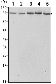
Figure 1: Western blot analysis using STAT3 mouse mAb against Hela (1),NIH/3T3 (2), Jurkat (3), PC-12 (4) and COS7 (5) cell lysate.
Immunohistochemical analysis

Figure 2: Immunohistochemical analysis of paraffin-embedded mammary cancer tissues (left) and lung cancer tissues (right) using STAT3 mouse mAb with DAB staining.
Immunofluorescence analysis

Figure 3: Immunofluorescence analysis of Hela cells using STAT3 mouse mAb (green). Blue: DRAQ5 fluorescent DNA dye. Red: Actin filaments have been labeled with Alexa Fluor-555 phalloidin.
For Research Use Only. Not for use in diagnostic procedures.