

SST Primary Antibody
Item Information
Catalog #
Size
Price
Description
The preproprotein encoded by this gene. Somatostatin is expressed throughout the body and inhibits the release of numerous secondary hormones by binding to high-affinity G-protein-coupled somatostatin receptors. This hormone is an important regulator of the endocrine system through its interactions with pituitary growth hormone, thyroid stimulating hormone, and most hormones of the gastrointestinal tract. Somatostatin also affects rates of neurotransmission in the central nervous system and proliferation of both normal and tumorigenic cells.
Product Overview
Entrez GenelD
6750
Aliases
SMST
Clone#
7G5
Host / Isotype
Mouse / IgG1
Species Reactivity
Human
Immunogen
Purified recombinant fragment of human SST (AA: 1-116) expressed in E. Coli.
Formulation
Purified antibody in PBS with 0.05% sodium azide
Storage
Store at 4°C short term. Aliquot and store at -20°C long term. Avoid freeze/thaw cycles.
Product Applications
WB (Western Blot)
1/500 - 1/2000
IHC_P(Immunohistochemistry)
1/200 - 1/1000
FCM (Flow Cytometry)
1/200 - 1/400
ELISA
1/10000
References
1.Acta Neurol Scand. 2010 Apr;121(4):225-9. 2.Endocrinology. 2009 May;150(5):2254-63.
Product Image
Western Blot
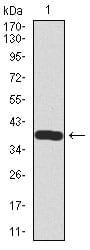
Figure 1: Western blot analysis using SST mAb against human SST recombinant protein. (Expected MW is 38.2 kDa)
Flow cytometric

Figure 2: Flow cytometric analysis of HepG2 cells using SST mouse mAb (green) and negative control (red).
Immunohistochemical analysis

Figure 3: Immunohistochemical analysis of paraffin-embedded pancreas tissues using SST mouse mAb with DAB staining.
Immunohistochemical analysis

Figure 4: Immunohistochemical analysis of paraffin-embedded lung cancer tissues using SST mouse mAb with DAB staining.
Elisa

Black line: Control Antigen (100 ng); Purple line: Antigen(10ng); Blue line: Antigen (50 ng); Red line: Antigen (100 ng);
For Research Use Only. Not for use in diagnostic procedures.