

SRA Primary Antibody
Item Information
Catalog #
Size
Price
Description
Steroid receptor RNA activator 1 (SRA), with 237-amino acid protein (about 27kDa), belongs to the growing family of functional non-coding RNAs. SRA was originally described as the first functional noncoding RNA able to specifically coactivate the activity of steroid receptors. Specifically, SRA exists as both an RNA transcript that forms a complex with steroid receptor coactivator-1 and as a stably expressed protein. Its expression is strongly up-regulated in many human tumors of the breast, uterus, and ovary, suggesting a potential role in pathogenesis. Although coactivation of steroid-dependent transcription by SRA is accompanied by a proliferative response, overexpression is not in itself sufficient to induce turmorigenesis.
Product Overview
Entrez GenelD
10011
Aliases
SRAP; STRAA1
Clone#
1D4H8
Host / Isotype
Mouse / IgG1
Species Reactivity
Human
Immunogen
Purified recombinant fragment of SRA expressed in E. Coli.
Formulation
Ascitic fluid containing 0.03% sodium azide.
Storage
Store at 4°C short term. Aliquot and store at -20°C long term. Avoid freeze/thaw cycles.
Product Applications
WB (Western Blot)
1/500 - 1/2000
IHC_P(Immunohistochemistry)
1/200 - 1/1000
ELISA
1/10000
References
1. Rainer B. Lanz, Steven S. Chua, Niall Barron. Mol. Cell. Biol, Oct 2003; 23: 7163 - 7176.
2. Shilpa Chooniedass-Kothari, Mohammad Kariminia Hamedani, Sandy Troup. Int J Cancer. 2006 Feb 15;118(4):1054-9
3. S. Chooniedass-Kothari, E. Emberley, M. K. Hamedani. FEBS Lett. 2004 May 21;566(1-3):43-7
2. Shilpa Chooniedass-Kothari, Mohammad Kariminia Hamedani, Sandy Troup. Int J Cancer. 2006 Feb 15;118(4):1054-9
3. S. Chooniedass-Kothari, E. Emberley, M. K. Hamedani. FEBS Lett. 2004 May 21;566(1-3):43-7
Product Image
Western Blot

Figure 1: Western blot analysis using SRA mouse mAb against truncated SRA recombinant protein (1), human ovary cancer tissue lysate (2) and A431 cell lysate (3).
Immunohistochemical analysis
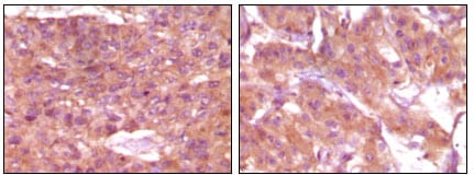
Figure 2: Immunohistochemical analysis of paraffin-embedded human skin carcinoma (left) and breast carcinoma (right), showing cytoplasmic and membrane localization using SRA mouse mAb with DAB staining.
For Research Use Only. Not for use in diagnostic procedures.