SORL1 Primary Antibody

Item Information
Catalog #
Size
Price
Description

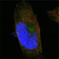
SORL1 (sortilin-related receptor, L A repeats containing) also known as sorting protein-related receptor containing LDLR class A (SorLA), is a Type I membrane protein that may be involved in cell-cell interaction. SorLA, a single transmembrane receptor, binds LDL and transports it into cells by endocytosis. SorLA is synthesized as a proreceptor which is processed to the mature form by a furin-like propeptidase. It can also bind to RAP (receptor-associated protein). SorLA is a multifunctional endocytis receptor important in lipoprotein and protease uptake. The N-terminal propeptide, which is removed, can be cleaved by furin or homologous proteases. Endogenous SorLA binds the neuropeptide head activator (HA) and is important for HA signaling and function. The gene encoding for the protein maps to chromosome 8p23.1. SorLA is expressed mainly in brain (cerebral cortex, cerebellum and the occipital pole), but can also be found in liver, spinal cord, kidney, testis and pancreas.

Product Overview
Entrez GenelD
6653
Aliases
SORL1
Clone#
3B6B11
Host / Isotype
Mouse / IgG1
Species Reactivity
Human
Immunogen
Purified recombinant fragment of human SORL1 expressed in E. Coli.
Formulation
Ascitic fluid containing 0.03% sodium azide.
Storage
4°C; -20°C for long term storage
Product Applications
WB (Western Blot)
1/500 - 1/2000
IHC_P(Immunohistochemistry)
1/200 - 1/1000
ICC (Immunocytochemistry)
1/200 - 1/1000
ELISA
1/10000
References
1. Scherzer CR. Offe K. Gearing M. et al. Arch Neurol. 2004, Aug, 61(8):1200-5.
2. Gabrielsson BG. Olofsson LE. Sjogren A. et al. Obes Res. 2005, Apr, 13(4):649-52.
3. Shah S. Yu G. Mol Interv. 2006, Apr, 6(2):74-6, 58. Review.
Product Image
Western Blot
Figure 1: Western blot analysis using SORL1 mouse mAb against truncated SORL1 recombinant protein.
Immunohistochemical analysis
Figure 2: Immunohistochemical analysis of paraffin-embedded human cerebrum tissues using SORL1 mouse mAb with DAB staining.
Immunohistochemical analysis
Figure 3: Immunohistochemical analysis of paraffin-embedded human brain, cortex tissues using SORL1 mouse mAb.
Immunofluorescence analysis
Figure 4: Confocal Immunofluorescence analysis of PANC-1 cells using SORL1 mouse mAb (green). Red: Actin filaments have been labeled with Alexa Fluor-555 phalloidin. Blue: DRAQ5 fluorescent DNA dye.
For Research Use Only. Not for use in diagnostic procedures.