

SOD2 Primary Antibody
Item Information
Catalog #
Size
Price
Description
This gene is a member of the iron/manganese superoxide dismutase family. It encodes a mitochondrial protein that forms a homotetramer and binds one manganese ion per subunit. This protein binds to the superoxide byproducts of oxidative phosphorylation and converts them to hydrogen peroxide and diatomic oxygen. Mutations in this gene have been associated with idiopathic cardiomyopathy (IDC), premature aging, sporadic motor neuron disease, and cancer. Alternative splicing of this gene results in multiple transcript variants. A related pseudogene has been identified on chromosome 1.
Product Overview
Entrez GenelD
6648
Aliases
IPOB; IPO-B; MNSOD; MVCD6; Mn-SOD
Clone#
8H3F9
Host / Isotype
Mouse / IgG1
Species Reactivity
Human
Immunogen
Purified recombinant fragment of human SOD2 (AA: 1-222) expressed in E. Coli.
Formulation
Purified antibody in PBS with 0.05% sodium azide
Storage
4°C; -20°C for long term storage
Product Applications
WB (Western Blot)
1/500 - 1/2000
IHC_P(Immunohistochemistry)
1/200 - 1/1000
FCM (Flow Cytometry)
1/200 - 1/400
ELISA
1/10000
References
1.Dis Markers. 2015;2015:746329.
2.Free Radic Biol Med. 2015 Dec;89:379-86.
2.Free Radic Biol Med. 2015 Dec;89:379-86.
Product Image
Elisa
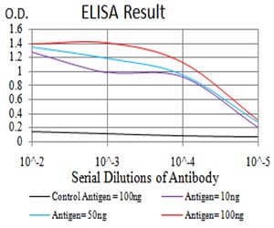
Figure 1: Black line: Control Antigen (100 ng);Purple line: Antigen (10ng); Blue line: Antigen (50 ng); Red line:Antigen (100 ng)
Western Blot
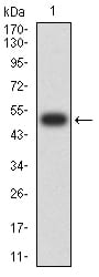
Figure 2:Western blot analysis using SOD2 mAb against human SOD2 (AA: 1-222) recombinant protein. (Expected MW is 50.7 kDa)
Western Blot
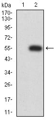
Figure 3:Western blot analysis using SOD2 mAb against HEK293 (1) and SOD2 (AA: 1-222)-hIgGFc transfected HEK293 (2) cell lysate.
Western Blot
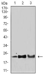
Figure 4:Western blot analysis using SOD2 mouse mAb against Hela (1), HepG2 (2), and SH-SY5Y (3) cell lysate.
Flow cytometric
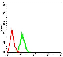
Figure 5:Flow cytometric analysis of MCF-7 cells using SOD2 mouse mAb (green) and negative control (red).
Immunohistochemical analysis
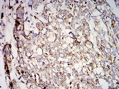
Figure 6:Immunohistochemical analysis of paraffin-embedded breast cancer tissues using SOD2 mouse mAb with DAB staining.
Immunohistochemical analysis
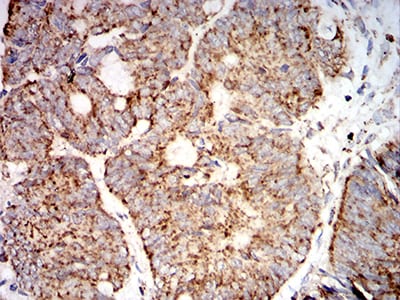
Figure 7:Immunohistochemical analysis of paraffin-embedded rectum cancer tissues using SOD2 mouse mAb with DAB staining.
For Research Use Only. Not for use in diagnostic procedures.