

SMCP Primary Antibody
Item Information
Catalog #
Size
Price
Description
Sperm mitochondria differ in morphology and subcellular localization from those of somatic cells. They are elongated, flattened, and arranged circumferentially to form a helical coiled sheath in the midpiece of the sperm flagellum. The protein encoded by this gene localizes to the capsule associated with the mitochondrial outer membranes and is thought to function in the organization and stabilization of the helical structure of the sperm's mitochondrial sheath.
Product Overview
Entrez GenelD
4184
Aliases
MCS; MCSP; HSMCSGEN1
Clone#
5C10D8
Host / Isotype
Mouse / IgG2b
Species Reactivity
Human
Immunogen
Purified recombinant fragment of human SMCP (AA: FULL(1-116)) expressed in E. Coli.
Formulation
Purified antibody from tissue culture in PBS with 0.05% sodium azide
Storage
Store at 4°C short term. Aliquot and store at -20°C long term. Avoid freeze/thaw cycles.
Product Applications
WB (Western Blot)
1/500 - 1/2000
ICC (Immunocytochemistry)
1/200 - 1/1000
ELISA
1/10000
References
1. Biol Reprod. 1999 Aug;61(2):428-35.
2. Mol Cell Biol. 2002 May;22(9):3046-52.
2. Mol Cell Biol. 2002 May;22(9):3046-52.
Product Image
Western Blot
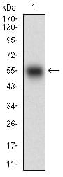
Figure 1: Western blot analysis using SMCP mAb against human SMCP (AA: FULL(1-116)) recombinant protein. (Expected MW is 38.3 kDa)
Western Blot
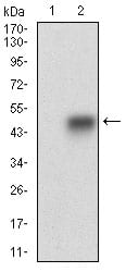
Figure 2: Western blot analysis using SMCP mAb against HEK293 (1) and SMCP (AA: FULL(1-116))-hIgGFc transfected HEK293 (2) cell lysate.
Immunofluorescence analysis
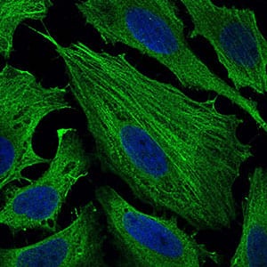
Figure 4: Immunofluorescence analysis of Hela cells using SMCP mouse mAb (green). Blue: DRAQ5 fluorescent DNA dye. Secondary antibody from Fisher (Cat#: 35503)
Elisa

Black line: Control Antigen (100 ng); Purple line: Antigen(10ng); Blue line: Antigen (50 ng); Red line: Antigen (100 ng);
For Research Use Only. Not for use in diagnostic procedures.