

SMARCA1 Primary Antibody
Item Information
Catalog #
Size
Price
Description
This gene encodes a member of the SWI/SNF family of proteins. The encoded protein is an ATPase which is expressed in diverse tissues and contributes to the chromatin remodeling complex that is involved in transcription. The protein may also play a role in DNA damage, growth inhibition and apoptosis of cancer cells. Alternative splicing results in multiple transcript variants.
Product Overview
Entrez GenelD
6594
Aliases
SWI; ISWI; SWI2; SNF2L; SNF2L1; SNF2LB; SNF2LT; hSNF2L; NURF140
Clone#
2H7B8
Host / Isotype
Mouse / IgG1
Species Reactivity
Human, Mouse
Immunogen
Purified recombinant fragment of human SMARCA1 (AA: 933-1070) expressed in E. Coli.
Formulation
Purified antibody in PBS with 0.05% sodium azide
Storage
Store at 4°C short term. Aliquot and store at -20°C long term. Avoid freeze/thaw cycles.
Product Applications
WB (Western Blot)
1/500 - 1/2000
IHC_P(Immunohistochemistry)
1/200 - 1/1000
FCM (Flow Cytometry)
1/200 - 1/400
ELISA
1/10000
References
1.Yonsei Med J. 2013 May 1;54(3):772-7.
2.BMC Med Genet. 2008 Feb 26;9:11.
2.BMC Med Genet. 2008 Feb 26;9:11.
Product Image
Elisa
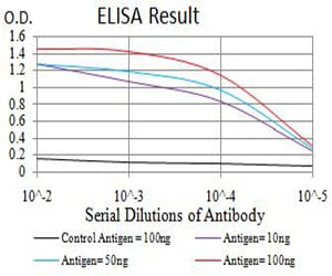
Figure 1: Black line: Control Antigen (100 ng);Purple line: Antigen (10ng); Blue line: Antigen (50 ng); Red line:Antigen (100 ng)
Western Blot
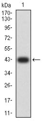
Figure 2:Western blot analysis using SMARCA1 mAb against human SMARCA1 (AA: 933-1070) recombinant protein. (Expected MW is 42.4 kDa)
Western Blot
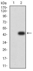
Figure 3:Western blot analysis using SMARCA1 mAb against HEK293 (1) and SMARCA1 (AA: 933-1070)-hIgGFc transfected HEK293 (2) cell lysate.
Western Blot
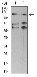
Figure 4:Western blot analysis using SMARCA1 mouse mAb against SW620 (1) and HT-29 (2) cell lysate.
Flow cytometric
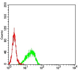
Figure 5:Flow cytometric analysis of NIH/3T3 cells using SMARCA1 mouse mAb (green) and negative control (red).
Immunohistochemical analysis
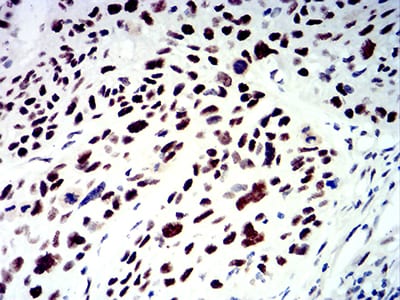
Figure 6:Immunohistochemical analysis of paraffin-embedded esophageal cancer tissues using SMARCA1 mouse mAb with DAB staining.
Immunohistochemical analysis
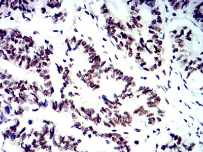
Figure 7:Immunohistochemical analysis of paraffin-embedded rectum cancer tissues using SMARCA1 mouse mAb with DAB staining.
For Research Use Only. Not for use in diagnostic procedures.