

SLC2A4 Primary Antibody
Item Information
Catalog #
Size
Price
Description
This gene is a member of the solute carrier family 2 (facilitated glucose transporter) family and encodes a protein that functions as an insulin-regulated facilitative glucose transporter. In the absence of insulin, this integral membrane protein is sequestered within the cells of muscle and adipose tissue. Within minutes of insulin stimulation, the protein moves to the cell surface and begins to transport glucose across the cell membrane. Mutations in this gene have been associated with noninsulin-dependent diabetes mellitus (NIDDM).
Product Overview
Entrez GenelD
6517
Aliases
GLUT4
Clone#
3G10A3
Host / Isotype
Mouse / IgG2b
Species Reactivity
Human, Mouse
Immunogen
Purified recombinant fragment of human SLC2A4 (AA: 224-353 ) expressed in E. Coli.
Formulation
Purified antibody in PBS with 0.05% sodium azide
Storage
Store at 4°C short term. Aliquot and store at -20°C long term. Avoid freeze/thaw cycles.
Product Applications
WB (Western Blot)
1/500 - 1/2000
IHC_P(Immunohistochemistry)
1/200 - 1/1000
ICC (Immunocytochemistry)
1/200 - 1/1000
FCM (Flow Cytometry)
1/200 - 1/400
ELISA
1/10000
References
1.J Biol Chem. 2011 May 13;286(19):16541-5.
2.PLoS One. 2010 Dec 20;5(12):e15560.
2.PLoS One. 2010 Dec 20;5(12):e15560.
Product Image
Western Blot

Figure 1: Western blot analysis using SLC2A4 mAb against human SLC2A4 recombinant protein. (Expected MW is 39.9 kDa)
Western Blot

Figure 2: Western blot analysis using SLC2A4 mAb against HEK293 (1) and SLC2A4 (AA: 224-353)-hIgGFc transfected HEK293 (2) cell lysate.
Immunofluorescence analysis
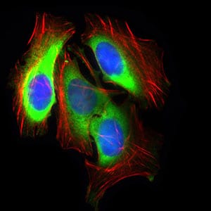
Figure 3: Immunofluorescence analysis of HeLa cells using SLC2A4 mouse mAb (green). Blue: DRAQ5 fluorescent DNA dye. Red: Actin filaments have been labeled with Alexa Fluor-555 phalloidin.
Immunofluorescence analysis

Figure 3: Immunofluorescence analysis of HepG2 cells using SLC2A4 mouse mAb (green). Blue: DRAQ5 fluorescent DNA dye. Red: Actin filaments have been labeled with Alexa Fluor-555 phalloidin.
Western Blot

Figure 3: Western blot analysis using SLC2A4 mouse mAb against HeLa (1), NIH3T3 (2), 3T3-L1 (3) cell lysate and Mouse heart (4) tissue lysate.
Flow cytometric

Figure 6: Flow cytometric analysis of HeLa cells using SLC2A4 mouse mAb (green) and negative control (purple).
Immunohistochemical analysis

Figure 7: Immunohistochemical analysis of paraffin-embedded bladder cancer tissues using SLC2A4 mouse mAb with DAB staining.
Immunohistochemical analysis

Figure 8: Immunohistochemical analysis of paraffin-embedded cardiac muscle tissues using SLC2A4 mouse mAb with DAB staining.
Elisa

Black line: Control Antigen (100 ng); Purple line: Antigen(10ng); Blue line: Antigen (50 ng); Red line: Antigen (100 ng);
For Research Use Only. Not for use in diagnostic procedures.