
SLC22A12 Primary Antibody
Item Information
Catalog #
Size
Price
Description
The protein encoded by this gene is a member of the organic anion transporter (OAT) family, and it acts as a urate transporter to regulate urate levels in blood. This protein is an integral membrane protein primarily found in epithelial cells of the proximal tubule of the kidney. An elevated level of serum urate, hyperuricemia, is associated with increased incidences of gout, and mutations in this gene cause renal hypouricemia type 1. Alternative splicing results in multiple transcript variants.
Product Overview
Entrez GenelD
116085
Aliases
RST; OAT4L; URAT1
Clone#
1B1G7
Host / Isotype
Mouse / Mouse IgG1
Species Reactivity
Human, Mouse
Immunogen
Purified recombinant fragment of human SLC22A12 (AA: 30-145) expressed in E. Coli.
Formulation
Purified antibody in PBS with 0.05% sodium azide
Storage
Store at 4°C short term. Aliquot and store at -20°C long term. Avoid freeze/thaw cycles.
Product Applications
WB (Western Blot)
1/500 - 1/2000
IHC_P(Immunohistochemistry)
1/200 - 1/1000
FCM (Flow Cytometry)
1/200 - 1/400
ELISA
1/10000
References
1.Rheumatology (Oxford). 2020 Dec 1;59(12):3988-3990.
2.PLoS One. 2020 Apr 9;15(4):e0231336.
2.PLoS One. 2020 Apr 9;15(4):e0231336.
Product Image
Elisa

Figure 1:Black line: Control Antigen (100 ng);Purple line: Antigen (10ng); Blue line: Antigen (50 ng); Red line:Antigen (100 ng)
Western Blot

Figure 2:Western blot analysis using SLC22A12 mAb against human SLC22A12 (AA: 30-145) recombinant protein. (Expected MW is 38.8 kDa)
Western Blot

Figure 3:Western blot analysis using SLC22A12 mAb against HEK293-6e (1) and SLC22A12 (AA: 30-145)-hIgGFc transfected HEK293-6e (2) cell lysate.
Western Blot

Figure 4:Western blot analysis using SLC22A12 mouse mAb against mouse kindey(1) tissue lysate.
Immunofluorescence analysis

Figure 5:Flow cytometric analysis of Hela cells using SLC22A12 mouse mAb (green) and negative control (red).
Immunofluorescence analysis
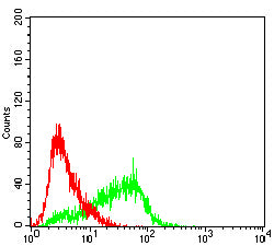
Figure 6:Flow cytometric analysis of HepG2 cells using SLC22A12 mouse mAb (green) and negative control (red).
Immunohistochemical analysis

Figure 7:Immunohistochemical analysis of paraffin-embedded kidney tissues using SLC22A12 mouse mAb with DAB staining.
For Research Use Only. Not for use in diagnostic procedures.