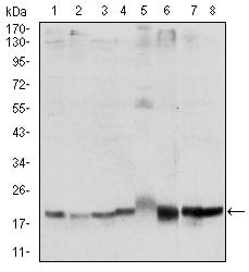
SKP1 Primary Antibody

Item Information
Catalog #
Size
Price
Description

This gene encodes a component of SCF complexes, which are composed of this protein, cullin 1, a ring-box protein, and one member of the F-box family of proteins. This protein binds directly to the F-box motif found in F-box proteins. SCF complexes are involved in the regulated ubiquitination of specific protein substrates, which targets them for degradation by the proteosome. Specific F-box proteins recognize different target protein(s), and many specific SCF substrates have been identified including regulators of cell cycle progression and development. Studies have also characterized the protein as an RNA polymerase II elongation factor. Alternative splicing of this gene results in two transcript variants. A related pseudogene has been identified on chromosome 7.

Product Overview
Entrez GenelD
6500
Aliases
OCP2; p19A; EMC19; SKP1A; OCP-II; TCEB1L; MGC34403
Clone#
4E11
Host / Isotype
Mouse / IgG1
Species Reactivity
Human
Immunogen
Purified recombinant fragment of human SKP1 expressed in E. Coli.
Formulation
Ascitic fluid containing 0.03% sodium azide.
Storage
4°C; -20°C for long term storage
Product Applications
WB (Western Blot)
1/500 - 1/2000
IHC_P(Immunohistochemistry)
1/200 - 1/1000
ICC (Immunocytochemistry)
1/200 - 1/1000
FCM (Flow Cytometry)
1/200 - 1/400
ELISA
1/10000
References
1. J Virol. 2009 Dec;83(23):12068-83.
2. Cell. 2009 Jul 23;138(2):389-403.
Product Image
Western Blot
Figure 1: Western blot analysis using SKP1 mAb against human SKP1 (AA: 1-160) recombinant protein. (Expected MW is 43.5 kDa)
Western Blot
Figure 2: Western blot analysis using SKP1 mouse mAb against Hela (1), NIH/3T3 (2), A431 (3), RAJI (4), PC-12 (5), Cos7 (6), MCF-7 (7) and HepG2 (8) cell lysate.
Immunohistochemical analysis
Figure 3: Immunohistochemical analysis of paraffin-embedded rectum cancer tissues using SKP1 mouse mAb with DAB staining.
Immunofluorescence analysis
Figure 4: Immunofluorescence analysis of Hela cells using SKP1 mouse mAb (green). Blue: DRAQ5 fluorescent DNA dye. Red: Actin filaments have been labeled with Alexa Fluor-555 phalloidin.
Flow cytometric
Figure 5: Flow cytometric analysis of NIH/3T3 cells using SKP1 mouse mAb (green) and negative control (red).
Elisa
Black line: Control Antigen (100 ng); Purple line: Antigen(10ng); Blue line: Antigen (50 ng); Red line: Antigen (100 ng);
For Research Use Only. Not for use in diagnostic procedures.