

SIRT7 Primary Antibody
Item Information
Catalog #
Size
Price
Description
This gene encodes a member of the sirtuin family of proteins, homologs to the yeast Sir2 protein. Members of the sirtuin family are characterized by a sirtuin core domain and grouped into four classes. The functions of human sirtuins have not yet been determined; however, yeast sirtuin proteins are known to regulate epigenetic gene silencing and suppress recombination of rDNA. Studies suggest that the human sirtuins may function as intracellular regulatory proteins with mono-ADP-ribosyltransferase activity. The protein encoded by this gene is included in class IV of the sirtuin family.
Product Overview
Entrez GenelD
51547
Aliases
SIR2L7
Clone#
1E2G10
Host / Isotype
Mouse / IgG1
Species Reactivity
Human
Immunogen
Purified recombinant fragment of human SIRT7 (AA: 1-105) expressed in E. Coli.
Formulation
Purified antibody in PBS with 0.05% sodium azide
Storage
Store at 4°C short term. Aliquot and store at -20°C long term. Avoid freeze/thaw cycles.
Product Applications
WB (Western Blot)
1/500 - 1/2000
IHC_P(Immunohistochemistry)
1/200 - 1/1000
ELISA
1/10000
References
1.Clin Cancer Res. 2014 Jul 1;20(13):3434-45.
2.Mol Cell Proteomics. 2014 Jan;13(1):73-83.
2.Mol Cell Proteomics. 2014 Jan;13(1):73-83.
Product Image
Elisa
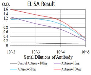
Figure 1: Black line: Control Antigen (100 ng); Purple line: Antigen(10ng); Blue line: Antigen (50 ng); Red line: Antigen (100 ng);
Western Blot

Figure 2:Western blot analysis using SIRT7 mAb against human SIRT7 (AA: 1-105) recombinant protein. (Expected MW is 38 kDa)
Western Blot

Figure 3:Western blot analysis using SIRT7 mAb against HEK293 (1) and SIRT7 (AA: 1-105)-hIgGFc transfected HEK293 (2) cell lysate.
Immunohistochemical analysis

Figure 4:Immunohistochemical analysis of paraffin-embedded liver cancer tissues using SIRT7 mouse mAb with DAB staining.
Immunohistochemical analysis

Figure 5:Immunohistochemical analysis of paraffin-embedded ovarian cancer tissues using SIRT7 mouse mAb with DAB staining.
For Research Use Only. Not for use in diagnostic procedures.