

SIRT4 Primary Antibody
Item Information
Catalog #
Size
Price
Description
This gene encodes a member of the sirtuin family of proteins, homologs to the yeast Sir2 protein. Members of the sirtuin family are characterized by a sirtuin core domain and grouped into four classes. The functions of human sirtuins have not yet been determined; however, yeast sirtuin proteins are known to regulate epigenetic gene silencing and suppress recombination of rDNA. Studies suggest that the human sirtuins may function as intracellular regulatory proteins with mono-ADP-ribosyltransferase activity. The protein encoded by this gene is included in class IV of the sirtuin family.
Product Overview
Entrez GenelD
23409
Aliases
SIR2L4
Clone#
6G6D2
Host / Isotype
Mouse / IgG1
Species Reactivity
Human
Immunogen
Purified recombinant fragment of human SIRT4 (AA: 215-314) expressed in E. Coli.
Formulation
Purified antibody in PBS with 0.05% sodium azide
Storage
Store at 4°C short term. Aliquot and store at -20°C long term. Avoid freeze/thaw cycles.
Product Applications
WB (Western Blot)
1/500 - 1/2000
ICC (Immunocytochemistry)
1/100 - 1/500
FCM (Flow Cytometry)
1/200 - 1/400
ELISA
1/10000
References
1.Eur J Histochem. 2011 Mar 21;55(1):e10.
2.J Biol Chem. 2014 Feb 14;289(7):4135-44.
2.J Biol Chem. 2014 Feb 14;289(7):4135-44.
Product Image
Elisa

Figure 1: Black line: Control Antigen (100 ng); Purple line: Antigen(10ng); Blue line: Antigen (50 ng); Red line: Antigen (100 ng);
Western Blot
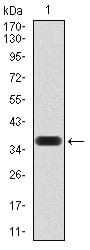
Figure 2:Western blot analysis using SIRT4 mAb against human SIRT4 (AA: 215-314) recombinant protein. (Expected MW is 37 kDa)
Western Blot

Figure 3:Western blot analysis using SIRT4 mAb against HEK293 (1) and SIRT4 (AA: 215-314)-hIgGFc transfected HEK293 (2) cell lysate.
Immunofluorescence analysis
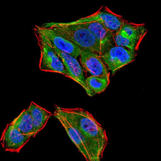
Figure 4:Immunofluorescence analysis of HeLa cells using SIRT4 mouse mAb (green). Blue: DRAQ5 fluorescent DNA dye. Red: Actin filaments have been labeled with Alexa Fluor- 555 phalloidin. Secondary antibody from Fisher (Cat#: 35503)
Flow cytometric

Figure 5:Flow cytometric analysis of HeLa cells using SIRT4 mouse mAb (green) and negative control (red).
For Research Use Only. Not for use in diagnostic procedures.