
Mouse Monoclonal Antibody to siglec15
Item Information
Catalog #
Size
Price
Description
SIGLEC15 (Sialic Acid Binding Ig Like Lectin 15) is a Protein Coding gene. Diseases associated with SIGLEC15 include Osteoporosis, Juvenile and Osteoporosis. Among its related pathways are Innate Immune System and RET signaling. An important paralog of this gene is SIGLEC1.
Product Overview
Entrez GenelD
284266
Aliases
CD33L3; HsT1361; SIGLEC-15
Clone#
3C9C3
Host / Isotype
Mouse / IgG1
Immunogen
Purified recombinant fragment of human Siglec15 (AA: Extra(20-263)) expressed in Mammal.
Formulation
Purified antibody in PBS with 0.05% sodium azide
Storage
Store at 4°C short term. Aliquot and store at -20°C long term. Avoid freeze/thaw cycles.
Product Applications
WB (Western Blot)
1/500 - 1/2000
IHC_P(Immunohistochemistry)
1/200 - 1/1000
ICC (Immunocytochemistry)
1/200 - 1/1000
FCM (Flow Cytometry)
1/200 - 1/400
ELISA
1/10000
References
1,J Biomed Sci. 2020 Jan 3;27(1):10.2,Glycobiology. 2013 Feb;23(2):178-87.
Product Image
Elisa
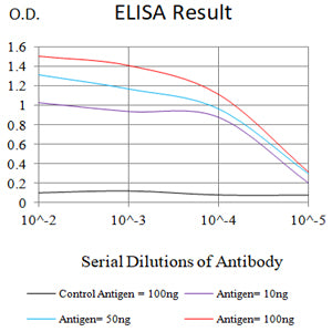
Figure 1:Black line: Control Antigen (100 ng);Purple line: Antigen (10ng); Blue line: Antigen (50 ng); Red line:Antigen (100 ng)
Western Blot
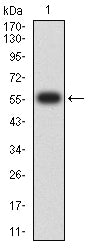
Figure 2:Western blot analysis using Siglec15 mAb against human Siglec15 (AA: Extra(20-263)) recombinant protein. (Expected MW is 56.4 kDa)
Flow cytometric analysis
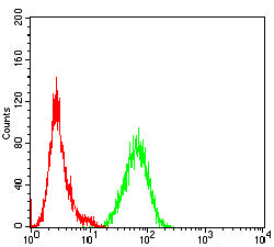
Figure 3:Flow cytometric analysis of Jurkat cells using Siglec15 mouse mAb (green) and negative control (red).
Flow cytometric analysis
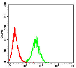
Figure 4:Flow cytometric analysis of THP-1 cells using Siglec15 mouse mAb (green) and negative control (red).
Immunohistochemical analysis
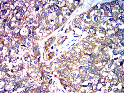
Figure 5:Immunohistochemical analysis of paraffin-embedded bladder cancer tissues using Siglec15 mouse mAb with DAB staining.
Western Blot
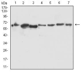
Figure 6:Western blot analysis using Siglec15 mouse mAb against PC-2 (1), LNCap (2), HEK293 (3), PC-3 (4), DU145 (5), COS-7 (6), and HEK293-6e (7) cell lysate.
Immunofluorescence analysis
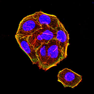
Figure 7:Immunofluorescence analysis of Hela cells using Siglec15 mouse mAb (green). Blue: DRAQ5 fluorescent DNA dye. Red: Actin filaments have been labeled with Alexa Fluor- 555 phalloidin. Secondary antibody from Fisher (Cat#: 35503)
For Research Use Only. Not for use in diagnostic procedures.