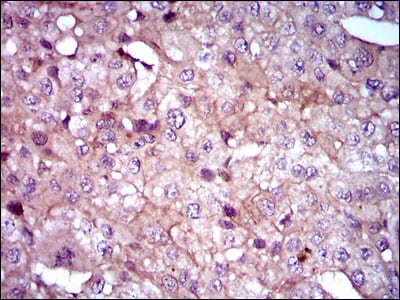
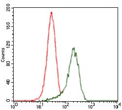
SHH Primary Antibody

Item Information
Catalog #
Size
Price
Description

This gene encodes a protein that is instrumental in patterning the early embryo. It has been implicated as the key inductive signal in patterning of the ventral neural tube, the anterior-posterior limb axis, and the ventral somites. Of three human proteins showing sequence and functional similarity to the sonic hedgehog protein of Drosophila, this protein is the most similar. The protein is made as a precursor that is autocatalytically cleaved; the N-terminal portion is soluble and contains the signalling activity while the C-terminal portion is involved in precursor processing. More importantly, the C-terminal product covalently attaches a cholesterol moiety to the N-terminal product, restricting the N-terminal product to the cell surface and preventing it from freely diffusing throughout the developing embryo. Defects in this protein or in its signalling pathway are a cause of holoprosencephaly (HPE), a disorder in which the developing forebrain fails to correctly separate into right and left hemispheres. HPE is manifested by facial deformities. It is also thought that mutations in this gene or in its signalling pathway may be responsible for VACTERL syndrome, which is characterized by vertebral defects, anal atresia, tracheoesophageal fistula with esophageal atresia, radial and renal dysplasia, cardiac anomalies, and limb abnormalities. Additionally, mutations in a long range enhancer located approximately 1 megabase upstream of this gene disrupt limb patterning and can result in preaxial polydactyly.

Product Overview
Entrez GenelD
6469
Aliases
TPT; HHG1; HLP3; HPE3; SMMCI; TPTPS; MCOPCB5
Clone#
5H4
Host / Isotype
Mouse / IgG1
Species Reactivity
Human, Mouse, Monkey
Immunogen
Purified recombinant fragment of human SHH (AA: 26-161) expressed in E. Coli.

Formulation
Purified antibody in PBS with 0.05% sodium azide
Storage
4°C; -20°C for long term storage
Product Applications
WB (Western Blot)
1/500 - 1/2000
IHC_P(Immunohistochemistry)
1/200 - 1/1000
FCM (Flow Cytometry)
1/200 - 1/400
ELISA
1/10000
References
1.Br J Cancer. 2010 Feb 16;102(4):738-47.
2.Mol Cancer. 2009 Dec 16;8:123.
Product Image
Western Blot
Figure 1: Western blot analysis using SHH mAb against human SHH recombinant protein. (Expected MW is 41 kDa)
Western Blot
Figure 2: Western blot analysis using SHH mAb against HEK293 (1) and SHH (AA: 26-161)-hIgGFc transfected HEK293 (2) cell lysate.
Western Blot
Figure 3: Western blot analysis using SHH mouse mAb against LNCaP (1), HepG2 (2), PANC-1 (3),HeLa (4), SK-N-SH (5), F9 (6), NIH3T3 (7), and COS7 (8) cell lysate.
Immunohistochemical analysis
Figure 4: Immunohistochemical analysis of paraffin-embedded liver cancer tissues using SHH mouse mAb with DAB staining.
Flow cytometric
Figure 5: Flow cytometric analysis of HeLa cells using SHH mouse mAb (green) and negative control (red).
Elisa
Black line: Control Antigen (100 ng); Purple line: Antigen(10ng); Blue line: Antigen (50 ng); Red line: Antigen (100 ng);
For Research Use Only. Not for use in diagnostic procedures.