

SHC1 Primary Antibody
Item Information
Catalog #
Size
Price
Description
This gene encodes three main isoforms that differ in activities and subcellular location. While all three are adapter proteins in signal transduction pathways, the longest (p66Shc) may be involved in regulating life span and the effects of reactive oxygen species. The other two isoforms, p52Shc and p46Shc, link activated receptor tyrosine kinases to the Ras pathway by recruitment of the GRB2/SOS complex. p66Shc is not involved in Ras activation. Unlike the other two isoforms, p46Shc is targeted to the mitochondrial matrix. Several transcript variants encoding different isoforms have been found for this gene.
Product Overview
Entrez GenelD
6464
Aliases
SHC; SHCA
Clone#
2F7C7
Host / Isotype
Mouse / IgG1
Species Reactivity
Human, Mouse
Immunogen
Purified recombinant fragment of human SHC1 (AA: 385-495) expressed in E. Coli.
Formulation
Purified antibody in PBS with 0.05% sodium azide.
Storage
Store at 4°C short term. Aliquot and store at -20°C long term. Avoid freeze/thaw cycles.
Product Applications
WB (Western Blot)
1/500 - 1/2000
ICC (Immunocytochemistry)
1/200 - 1/1000
FCM (Flow Cytometry)
1/200 - 1/400
ELISA
1/10000
References
1. Am J Physiol Heart Circ Physiol. 2012 Feb 1;302(3):H724-32.
2. Clin Cardiol. 2010 Sep;33(9):548-52.
2. Clin Cardiol. 2010 Sep;33(9):548-52.
Product Image
Western Blot

Figure 1: Western blot analysis using SHC1 mAb against human SHC1 (AA: 385-495) recombinant protein. (Expected MW is 37.3 kDa)
Western Blot

Figure 2: Western blot analysis using SHC1 mAb against HEK293 (1) and SHC1 (AA: 385-495)-hIgGFc transfected HEK293 (2) cell lysate.
Western Blot

Figure 3: Western blot analysis using SHC1 mouse mAb against NIH/3T3 cell lysate.
Immunofluorescence analysis
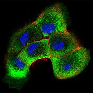
Figure 4: Immunofluorescence analysis of A431 cells using SHC1 mouse mAb (green). Blue: DRAQ5 fluorescent DNA dye. Red: Actin filaments have been labeled with Alexa Fluor-555 phalloidin. Secondary antibody from Fisher (Cat#: 35503)
Flow cytometric

Figure 5: Flow cytometric analysis of NIH/3T3 cells using SHC1 mouse mAb (green) and negative control (red).
Elisa

Black line: Control Antigen (100 ng); Purple line: Antigen(10ng); Blue line: Antigen (50 ng); Red line: Antigen (100 ng);
For Research Use Only. Not for use in diagnostic procedures.