

SH3GL1 Primary Antibody
Item Information
Catalog #
Size
Price
Description
This gene encodes a member of the endophilin family of Src homology 3 domain-containing proteins. The encoded protein is involved in endocytosis and may also play a role in the cell cycle. Overexpression of this gene may play a role in leukemogenesis, and the encoded protein has been implicated in acute myeloid leukemia as a fusion partner of the myeloid-lymphoid leukemia protein. Pseudogenes of this gene are located on the long arm of chromosomes 11 and 17. Alternatively spliced transcript variants encoding multiple isoforms have been observed for this gene.
Product Overview
Entrez GenelD
6455
Aliases
EEN; CNSA1; SH3P8; SH3D2B
Clone#
2A9H4
Host / Isotype
Mouse / IgG1
Species Reactivity
Human
Immunogen
Purified recombinant fragment of human SH3GL1 (AA: 12-119) expressed in E. Coli.
Formulation
Purified antibody in PBS with 0.05% sodium azide
Storage
Store at 4°C short term. Aliquot and store at -20°C long term. Avoid freeze/thaw cycles.
Product Applications
WB (Western Blot)
1/500 - 1/2000
FCM (Flow Cytometry)
1/200 - 1/400
ELISA
1/10000
References
1.J Exp Clin Cancer Res. 2012 Oct 11;31:85.
2.Zhonghua Wai Ke Za Zhi. 2010 Mar 15;48(6):435-8.
2.Zhonghua Wai Ke Za Zhi. 2010 Mar 15;48(6):435-8.
Product Image
Elisa
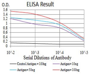
Figure 1: Black line: Control Antigen (100 ng);Purple line: Antigen (10ng); Blue line: Antigen (50 ng); Red line:Antigen (100 ng)
Western Blot
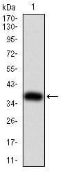
Figure 2:Western blot analysis using SH3GL1 mAb against human SH3GL1 (AA: 12-119) recombinant protein. (Expected MW is 37.6 kDa)
Western Blot
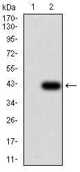
Figure 3:Western blot analysis using SH3GL1 mAb against HEK293 (1) and SH3GL1 (AA: 12-119)-hIgGFc transfected HEK293 (2) cell lysate.
Flow cytometric
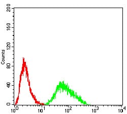
Figure 4:Flow cytometric analysis of Hela cells using SH3GL1 mouse mAb (green) and negative control (red).
For Research Use Only. Not for use in diagnostic procedures.