
SERPINE1 Primary Antibody
Item Information
Catalog #
Size
Price
Description
This gene encodes a member of the serine proteinase inhibitor (serpin) superfamily. This member is the principal inhibitor of tissue plasminogen activator (tPA) and urokinase (uPA), and hence is an inhibitor of fibrinolysis. Defects in this gene are the cause of plasminogen activator inhibitor-1 deficiency (PAI-1 deficiency), and high concentrations of the gene product are associated with thrombophilia. Alternatively spliced transcript variants encoding different isoforms have been found for this gene.
Product Overview
Entrez GenelD
5054
Aliases
PAI; PAI1; PAI-1; PLANH1
Clone#
1D5
Host / Isotype
Mouse / IgG1
Species Reactivity
Human
Immunogen
Purified recombinant fragment of human SERPINE1 expressed in E. Coli.
Formulation
Purified antibody in PBS with 0.05% sodium azide
Storage
Store at 4°C short term. Aliquot and store at -20°C long term. Avoid freeze/thaw cycles.
Product Applications
WB (Western Blot)
1/500 - 1/2000
IHC_P(Immunohistochemistry)
1/200 - 1/1000
FCM (Flow Cytometry)
1/200 - 1/400
ELISA
1/10000
References
1. Biol Pharm Bull. 2009 Apr;32(4):573-7.
2. Clin Chim Acta. 2009 Apr;402(1-2):189-92.
2. Clin Chim Acta. 2009 Apr;402(1-2):189-92.
Product Image
Western Blot

Figure 1: Western blot analysis using SERPINE1 mAb against human SERPINE1 (AA: 194-316) recombinant protein. (Expected MW is 45kDa kDa)
Western Blot

Figure 2: Western blot analysis using SERPINE1 mAb against HEK293 (1) and SERPINE1 (AA: 194-316)-hIgGFc transfected HEK293 (2) cell lysate.
Immunohistochemical analysis

Figure 3: Immunohistochemical analysis of paraffin-embedded lung cancer tissues using SERPINE1 mouse mAb with DAB staining.
Immunohistochemical analysis
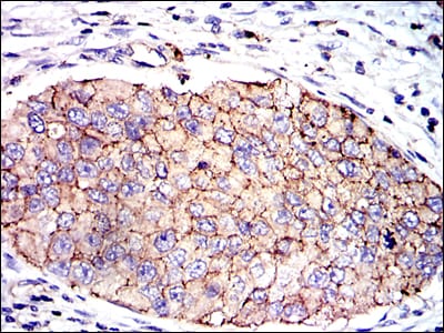
Figure 4: Immunohistochemical analysis of paraffin-embedded kidney cancer tissues using SERPINE1 mouse mAb with DAB staining.
Flow cytometric

Figure 5: Flow cytometric analysis of NIH/3T3 cells using SERPINE1 mouse mAb (green) and negative control (red).
Elisa

Black line: Control Antigen (100 ng); Purple line: Antigen(10ng); Blue line: Antigen (50 ng); Red line: Antigen (100 ng);
For Research Use Only. Not for use in diagnostic procedures.