

SERPINA3 Primary Antibody
Item Information
Catalog #
Size
Price
Description
The protein encoded by this gene is a plasma protease inhibitor and member of the serine protease inhibitor class. Polymorphisms in this protein appear to be tissue specific and influence protease targeting. Variations in this protein's sequence have been implicated in Alzheimer's disease, and deficiency of this protein has been associated with liver disease. Mutations have been identified in patients with Parkinson disease and chronic obstructive pulmonary disease.
Product Overview
Entrez GenelD
12
Aliases
ACT; AACT; GIG24; GIG25
Clone#
5G3C11
Host / Isotype
Mouse / IgG1
Species Reactivity
Human
Immunogen
Purified recombinant fragment of human SERPINA3 (AA: 279-432) expressed in E. Coli.
Formulation
Purified antibody in PBS with 0.05% sodium azide.
Storage
Store at 4°C short term. Aliquot and store at -20°C long term. Avoid freeze/thaw cycles.
Product Applications
WB (Western Blot)
1/500 - 1/2000
IHC_P(Immunohistochemistry)
1/200 - 1/1000
FCM (Flow Cytometry)
1/200 - 1/400
ELISA
1/10000
References
1. Cerebrovasc Dis. 2010;29(1):68-72.
2. Cerebrovasc Dis. 2007;23(1):46-9.
2. Cerebrovasc Dis. 2007;23(1):46-9.
Product Image
Western Blot

Figure 1: Western blot analysis using SERPINA3 mAb against human SERPINA3 (AA: 279-432) recombinant protein. (Expected MW is 42.3 kDa)
Western Blot

Figure 2: Western blot analysis using SERPINA3 mAb against HEK293 (1) and SERPINA3 (AA: 279-432)-hIgGFc transfected HEK293 (2) cell lysate.
Western Blot
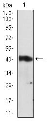
Figure 3: Western blot analysis using SERPINA3 mouse mAb against A549 cell lysate.
Flow cytometric

Figure 4: Flow cytometric analysis of A549 cells using SERPINA3 mouse mAb (green) and negative control (red).
Immunohistochemical analysis

Figure 5: Immunohistochemical analysis of paraffin-embedded endometrial cancer tissues using SERPINA3 mouse mAb with DAB staining.
Immunohistochemical analysis
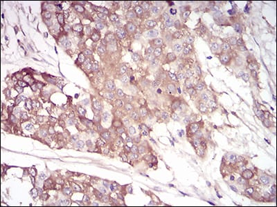
Figure 6: Immunohistochemical analysis of paraffin-embedded bladder cancer tissues using SERPINA3 mouse mAb with DAB staining.
Elisa

Black line: Control Antigen (100 ng); Purple line: Antigen(10ng); Blue line: Antigen (50 ng); Red line: Antigen (100 ng);
For Research Use Only. Not for use in diagnostic procedures.