

SERPINA Primary Antibody
Item Information
Catalog #
Size
Price
Description
The protein encoded by this gene is a serine protease inhibitor belonging to the serpin superfamily whose targets include elastase, plasmin, thrombin, trypsin, chymotrypsin, and plasminogen activator. This protein is produced in the liver, the bone marrow, by lymphocytic and monocytic cells in lymphoid tissue, and by the Paneth cells of the gut. Defects in this gene are associated with chronic obstructive pulmonary disease, emphysema, and chronic liver disease. Several transcript variants encoding the same protein have been found for this gene. [provided by RefSeq, Aug 2020]
Product Overview
Entrez GenelD
5265
Aliases
PI; A1A; AAT; PI1; A1AT; nNIF; PRO2275; alpha1AT
Clone#
7D3A2
Host / Isotype
Mouse / Mouse IgG1
Immunogen
Purified recombinant fragment of human SERPINA (AA: 269-419) expressed in E. Coli.
Formulation
Purified antibody in PBS with 0.05% sodium azide
Storage
Store at 4°C short term. Aliquot and store at -20°C long term. Avoid freeze/thaw cycles.
Product Applications
WB (Western Blot)
1/500 - 1/2000
IHC_P(Immunohistochemistry)
1/200-1/1000
FCM (Flow Cytometry)
1/200-1/400
ELISA
1/10000
References
1,Rev Med Virol. 2020 Aug 26;e2157.2,Clin Liver Dis. 2020 Aug;24(3):483-492.
Product Image
ELISA

Figure 1: Black line: Control Antigen (100 ng);Purple line: Antigen (10ng); Blue line: Antigen (50 ng); Red line: Antigen (100 ng)
WESTERN BLOT
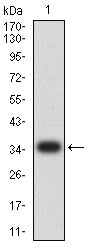
Figure 2: Western blot analysis using SERPINA mAb against human SERPINA (AA: 269-419) recombinant protein. (Expected MW is 35.7 kDa)
WESTERN BLOT
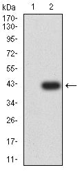
Figure 3: Western blot analysis using SERPINA mAb against HEK293-6e (1) and SERPINA (AA: 269-419)-hIgGFc transfected HEK293-6e (2) cell lysate.
FLOW CYTOMETRY

Figure 4: Flow cytometric analysis of Hela cells using SERPINA mouse mAb (green) and negative control (red).
FLOW CYTOMETRY

Figure 5: Flow cytometric analysis of Jurkat cells using SERPINA mouse mAb (green) and negative control (red).
IMMUNOHISTOCHEMISTRY
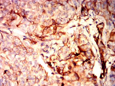
Figure 6: Immunohistochemical analysis of paraffin-embedded bladder cancer tissues using SERPINA mouse mAb with DAB staining.
IMMUNOHISTOCHEMISTRY
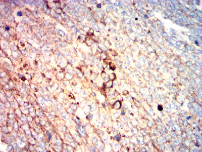
Figure 7: Immunohistochemical analysis of paraffin-embedded ovarian cancer tissues using SERPINA mouse mAb with DAB staining.
For Research Use Only. Not for use in diagnostic procedures.