

SELL Primary Antibody
Item Information
Catalog #
Size
Price
Description
This gene encodes a cell surface adhesion molecule that belongs to a family of adhesion/homing receptors. The encoded protein contains a C-type lectin-like domain, a calcium-binding epidermal growth factor-like domain, and two short complement-like repeats. The gene product is required for binding and subsequent rolling of leucocytes on endothelial cells, facilitating their migration into secondary lymphoid organs and inflammation sites. Single-nucleotide polymorphisms in this gene have been associated with various diseases including immunoglobulin A nephropathy. Alternatively spliced transcript variants have been found for this gene.
Product Overview
Entrez GenelD
6402
Aliases
TQ1; LAM1; LEU8; LNHR; LSEL; CD62L; LYAM1; PLNHR; LECAM1
Clone#
8C8B7
Host / Isotype
Mouse / IgG1
Species Reactivity
Human
Immunogen
Purified recombinant fragment of human SELL (AA: 83-186) expressed in E. Coli.
Formulation
Purified antibody in PBS with 0.05% sodium azide
Storage
Store at 4°C short term. Aliquot and store at -20°C long term. Avoid freeze/thaw cycles.
Product Applications
WB (Western Blot)
1/500 - 1/2000
ICC (Immunocytochemistry)
1/200 - 1/1000
ELISA
1/10000
References
PLoS One. 2012;7(9):e44814.
Eur J Cell Biol. 2012 Apr;91(4):257-64.
Eur J Cell Biol. 2012 Apr;91(4):257-64.
Product Image
Elisa
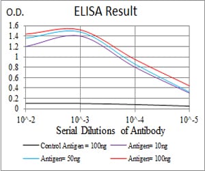
Figure 1: Black line: Control Antigen (100 ng); Purple line: Antigen(10ng); Blue line: Antigen (50 ng); Red line: Antigen (100 ng);
Western Blot

Figure 2:Western blot analysis using SELL mAb against human SELL (AA: 83-186) recombinant protein. (Expected MW is 37.4 kDa)
Western Blot

Figure 3:Western blot analysis using SELL mAb against HEK293 (1) and SELL (AA:83-186)-hIgGFc transfected HEK293 (2) cell lysate.
Immunofluorescence analysis

Figure 4:Immunofluorescence analysis of Hela cells using SELL mouse mAb (green). Blue: DRAQ5 fluorescent DNA dye. Red: Actin filaments have been labeled with Alexa Fluor- 555 phalloidin. Secondary antibody from Fisher (Cat#: 35503)
Immunofluorescence analysis

Figure 5:Immunofluorescence analysis of MCF-7 cells using SELL mouse mAb (green). Blue: DRAQ5 fluorescent DNA dye. Red: Actin filaments have been labeled with Alexa Fluor- 555 phalloidin. Secondary antibody from Fisher (Cat#: 35503)
For Research Use Only. Not for use in diagnostic procedures.