

SEC31A Primary Antibody
Item Information
Catalog #
Size
Price
Description
The protein encoded by this gene shares similarity with the yeast Sec31 protein, and is a component of the outer layer of the coat protein complex II (COPII). The encoded protein is involved in vesicle budding from the endoplasmic reticulum (ER) and contains multiple WD repeats near the N-terminus and a proline-rich region in the C-terminal half. It associates with the protein encoded by the SEC13 homolog, nuclear pore and COPII coat complex component (SEC13), and is required for ER-Golgi transport. Monoubiquitylation of this protein by CUL3-KLHL12 was found to regulate the size of COPII coats to accommodate unusually shaped cargo. Alternative splicing results in multiple transcript variants encoding different isoforms.
Product Overview
Entrez GenelD
22872
Aliases
ABP125; ABP130; HSPC275; HSPC334; SEC31L1
Clone#
5D12C6
Host / Isotype
Mouse / IgG1
Species Reactivity
Human
Immunogen
Purified recombinant fragment of human SEC31A (AA: 429-571) expressed in E. Coli.
Formulation
Purified antibody in PBS with 0.05% sodium azide
Storage
Store at 4°C short term. Aliquot and store at -20°C long term. Avoid freeze/thaw cycles.
Product Applications
WB (Western Blot)
1/500 - 1/2000
IHC_P(Immunohistochemistry)
1/200 - 1/1000
ELISA
1/10000
References
1.J Biol Chem. 2015 Feb 20;290(8):4981-93.
2.Blood. 2011 Apr 14;117(15):4056-64.
2.Blood. 2011 Apr 14;117(15):4056-64.
Product Image
Elisa
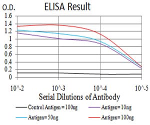
Figure 1: Black line: Control Antigen (100 ng);Purple line: Antigen (10ng); Blue line: Antigen (50 ng); Red line:Antigen (100 ng)
Western Blot
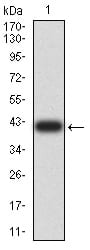
Figure 2:Western blot analysis using SEC31A mAb against human SEC31A (AA: 429-571) recombinant protein. (Expected MW is 41.8 kDa)
Western Blot
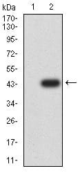
Figure 3:Western blot analysis using SEC31A mAb against HEK293 (1) and SEC31A (AA: 429-571)-hIgGFc transfected HEK293 (2) cell lysate.
Immunohistochemical analysis

Figure 4:Immunohistochemical analysis of paraffin-embedded ovarian cancer tissues using SEC31A mouse mAb with DAB staining.
For Research Use Only. Not for use in diagnostic procedures.