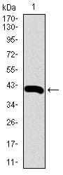
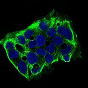
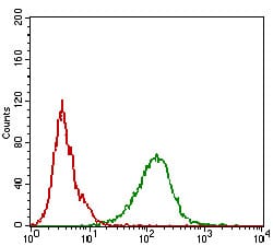
SDC1 Primary Antibody

Item Information
Catalog #
Size
Price
Description

The protein encoded by this gene is a transmembrane (type I) heparan sulfate proteoglycan and is a member of the syndecan proteoglycan family. The syndecans mediate cell binding, cell signaling, and cytoskeletal organization and syndecan receptors are required for internalization of the HIV-1 tat protein. The syndecan-1 protein functions as an integral membrane protein and participates in cell proliferation, cell migration and cell-matrix interactions via its receptor for extracellular matrix proteins. Altered syndecan-1 expression has been detected in several different tumor types. While several transcript variants may exist for this gene, the full-length natures of only two have been described to date. These two represent the major variants of this gene and encode the same protein.

Product Overview
Entrez GenelD
6382
Aliases
SDC; CD138; SYND1; syndecan
Clone#
1A3A3
Host / Isotype
Mouse / IgG1
Species Reactivity
Human, Mouse
Immunogen
Purified recombinant fragment of human SDC1 (AA: 28-171) expressed in E. Coli.
Formulation
Purified antibody in PBS with 0.05% sodium azide.
Storage
4°C; -20°C for long term storage
Product Applications
WB (Western Blot)
1/500 - 1/2000
ICC (Immunocytochemistry)
1/200 - 1/1000
FCM (Flow Cytometry)
1/200 - 1/400
ELISA
1/10000
References
1. Am J Clin Pathol. 2012 Mar;137(3):423-8.
2. PLoS One. 2011;6(9):e25252.
Product Image
Western Blot
Figure 1: Western blot analysis using SDC1 mAb against human SDC1 (AA: 28-171) recombinant protein. (Expected MW is 44.4 kDa)
Western Blot
Figure 2: Western blot analysis using SDC1 mAb against HEK293 (1) and SDC1 (AA: 28-171)-hIgGFc transfected HEK293 (2) cell lysate.
Western Blot
Figure 3: Western blot analysis using SDC1 mouse mAb against Hela (1), MCF-7 (2), HepG2 (3), T47D (4), Jurkat (5), NIH/3T3 (6) cell lysate.
Immunofluorescence analysis
Figure 4: Immunofluorescence analysis of HepG2 cells using SDC1 mouse mAb (green). Blue: DRAQ5 fluorescent DNA dye. Secondary antibody from Fisher (Cat#: 35503)
Flow cytometric
Figure 5: Flow cytometric analysis of A431 cells using SDC1 mouse mAb (green) and negative control (red).
Elisa
Black line: Control Antigen (100 ng); Purple line: Antigen(10ng); Blue line: Antigen (50 ng); Red line: Antigen (100 ng);
For Research Use Only. Not for use in diagnostic procedures.