

SCGB2A2 Primary Antibody
Item Information
Catalog #
Size
Price
Description
Mammaglobin is a gene that is expressed almost exclusively in the normal breast epithelium and human breast cancer. It is a member of the secretoglobin gene family and forms a heterodimer with lipophilin B. It has been suggested that mammaglobin may be a useful marker for breast cancer clinical research. Studies investigating the detection of mRNA by RT PCR from circulating carcinoma cells in the peripheral blood of breast cancer patients have shown that mammaglobin is a highly specific marker and correlates with several prognostic factors, such as lymph node involvement.Tissue specificity: Mammary gland specific. Over-expressed in breast cancer.
Product Overview
Entrez GenelD
4250
Aliases
MGB1; UGB2; MGC71974; SCGB2A2
Clone#
3C8
Host / Isotype
Mouse / IgG1
Species Reactivity
Human
Immunogen
Purified recombinant fragment of human SCGB2A2 expressed in E. Coli.
Formulation
Ascitic fluid containing 0.03% sodium azide.
Storage
Store at 4°C short term. Aliquot and store at -20°C long term. Avoid freeze/thaw cycles.
Product Applications
WB (Western Blot)
1/500 - 1/2000
IHC_P(Immunohistochemistry)
1/200 - 1/1000
ICC (Immunocytochemistry)
1/200 - 1/1000
FCM (Flow Cytometry)
1/200 - 1/400
ELISA
1/10000
References
1. Ann Oncol. 2006 Jun;17 Suppl 7:vii41-5.
2. Mod Pathol. 2007 Feb;20(2):208-14.
2. Mod Pathol. 2007 Feb;20(2):208-14.
Product Image
Western Blot

Figure 1: Western blot analysis using SCGB2A2 mAb against human SCGB2A2 (AA: 2-93) recombinant protein. (Expected MW is 35.8 kDa)
Immunohistochemical analysis

Figure 2: Immunohistochemical analysis of paraffin-embedded mammary cancer tissues using SCGB2A2 mouse mAb with DAB staining.
Immunofluorescence analysis

Figure 3: Immunofluorescence analysis of Hela cells using SCGB2A2 mouse mAb (green). Blue: DRAQ5 fluorescent DNA dye. Red: Actin filaments have been labeled with Alexa Fluor-555 phalloidin.
Flow cytometric
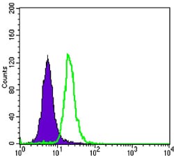
Figure 4: Flow cytometric analysis of SK-BR-3 cells using SCGB2A2 mouse mAb (green) and negative control (purple).
For Research Use Only. Not for use in diagnostic procedures.