

SATB2 Primary Antibody
Item Information
Catalog #
Size
Price
Description
This gene encodes a DNA binding protein that specifically binds nuclear matrix attachment regions. The encoded protein is involved in transcription regulation and chromatin remodeling. Defects in this gene are associated with isolated cleft palate and cognitive disability. Alternate splicing results in multiple transcript variants that encode the same protein. [provided by RefSeq, Feb 2010]
Product Overview
Entrez GenelD
23314
Aliases
GLSS
Clone#
7B2E11
Host / Isotype
Mouse / IgG1
Species Reactivity
Human
Immunogen
Purified recombinant fragment of human SATB2 (AA: 377-499) expressed in E. Coli.
Formulation
Purified antibody in PBS with 0.05% sodium azide
Storage
Store at 4°C short term. Aliquot and store at -20°C long term. Avoid freeze/thaw cycles.
Product Applications
WB (Western Blot)
1/500 - 1/2000
IHC_P(Immunohistochemistry)
1/200 - 1/1000
FCM (Flow Cytometry)
1/200 - 1/400
ELISA
1/10000
References
1.Lung Cancer. 2016 Aug;98:122-129.
2.Arch Dermatol Res. 2016 Aug;308(6):449-54.
2.Arch Dermatol Res. 2016 Aug;308(6):449-54.
Product Image
Elisa
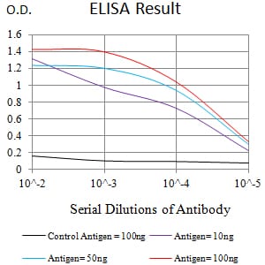
Figure 1:Black line: Control Antigen (100 ng);Purple line: Antigen (10ng); Blue line: Antigen (50 ng); Red line:Antigen (100 ng)
Western Blot

Figure 2:Western blot analysis using SATB2 mAb against human SATB2 (AA: 377-499) recombinant protein. (Expected MW is 39.7 kDa)
Western Blot
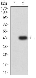
Figure 3:Western blot analysis using SATB2 mAb against HEK293 (1) and SATB2 (AA: 377-499)-hIgGFc transfected HEK293 (2) cell lysate.
Flow cytometric
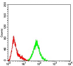
Figure 4:Flow cytometric analysis of Hela cells using SATB2 mouse mAb (green) and negative control (red).
Immunohistochemical analysis

Figure 5:Immunohistochemical analysis of paraffin-embedded rectum cancer tissues using SATB2 mouse mAb with DAB staining.
For Research Use Only. Not for use in diagnostic procedures.