
S100A10/P11 Primary Antibody
Item Information
Catalog #
Size
Price
Description
S100 calcium binding protein A10 (S100A10/P11), it is a member of the S100 family of proteins containing 2 EF-hand calcium-binding motifs. S100 proteins are localized in the cytoplasm and/or nucleus of a wide range of cells, and involved in the regulation of a number of cellular processes such as cell cycle progression and differentiation. S100 genes include at least 13 members which are located as a cluster on chromosome 1q21. This protein may function in exocytosis and endocytosis.
Product Overview
Entrez GenelD
6281
Aliases
S100A10; P11; PP11; PRSS26
Clone#
4E7E10
Host / Isotype
Mouse / IgG1
Species Reactivity
Human
Immunogen
Purified recombinant fragment of human P11 expressed in E. Coli.
Formulation
Ascitic fluid containing 0.03% sodium azide.
Storage
Store at 4°C short term. Aliquot and store at -20°C long term. Avoid freeze/thaw cycles.
Product Applications
WB (Western Blot)
1/500 - 1/2000
IHC_P(Immunohistochemistry)
1/200 - 1/1000
ICC (Immunocytochemistry)
1/200 - 1/1000
ELISA
1/10000
References
1. Svenningsson P, Greengard P. Curr Opin Pharmacol. 2007;7(1):27-32.
2. Santamaria-Kisiel L, Rintala-Dempsey AC, Shaw GS. Biochem J. 2006;396(2):201-14.
3. Rust R, VL, van dJ, Harms G,.et al. Br J Haematol. 2005;131(5):596-608.
2. Santamaria-Kisiel L, Rintala-Dempsey AC, Shaw GS. Biochem J. 2006;396(2):201-14.
3. Rust R, VL, van dJ, Harms G,.et al. Br J Haematol. 2005;131(5):596-608.
Product Image
Western Blot

Figure 1: Western blot analysis using S100A10/P11 mouse mAb against MCF-7 (1), HepG2 (2) and Hela (3).
Immunohistochemical analysis

Figure 2: Immunohistochemical analysis of paraffin-embedded human nerve and ganglion cells using S100A10/P11 mouse mAb.
Immunofluorescence analysis
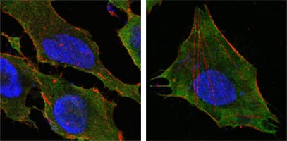
Figure 3: Confocal Immunofluorescence analysis of Hela (left) and L-02 (right) cells using S100A10/P11 mouse mAb(green). Red: Actin filaments have been labeled with DY-554 phalloidin. Blue: DRAQ5 fluorescent DNA dye.
For Research Use Only. Not for use in diagnostic procedures.