

RUNX3 Primary Antibody
Item Information
Catalog #
Size
Price
Description
This gene encodes a member of the runt domain-containing family of transcription factors. A heterodimer of this protein and a beta subunit forms a complex that binds to the core DNA sequence 5'-PYGPYGGT-3' found in a number of enhancers and promoters, and can either activate or suppress transcription. It also interacts with other transcription factors. It functions as a tumor suppressor, and the gene is frequently deleted or transcriptionally silenced in cancer. Multiple transcript variants encoding different isoforms have been found for this gene.
Product Overview
Entrez GenelD
864
Aliases
AML2; CBFA3; PEBP2aC; FLJ34510; MGC16070
Clone#
2B3
Host / Isotype
Mouse / IgG2b
Species Reactivity
Human, Mouse
Immunogen
Purified recombinant fragment of human RUNX3 (AA:186-252) expressed in E. Coli.
Formulation
Purified antibody in PBS with 0.05% sodium azide
Storage
Store at 4°C short term. Aliquot and store at -20°C long term. Avoid freeze/thaw cycles.
Product Applications
WB (Western Blot)
1/500 - 1/2000
IHC_P(Immunohistochemistry)
1/200 - 1/1000
ICC (Immunocytochemistry)
1/200 - 1/1000
FCM (Flow Cytometry)
1/200 - 1/400
ELISA
1/10000
References
1.J Cancer Res Clin Oncol. 2011 Dec;137(12):1823-30. 2.Oncogene. 2012 Jan 26;31(4):527-34.
Product Image
Western Blot

Figure 1: Western blot analysis using RUNX3 mAb against human RUNX3 recombinant protein. (Expected MW is 33 kDa)
Western Blot

Figure 2: Western blot analysis using RUNX3 mAb against HEK293 (1) and RUNX3 (AA: 186-252)-hIgGFc transfected HEK293 (2) cell lysate.
Flow cytometric

Figure 4: Flow cytometric analysis of NIH3T3 cells using RUNX3 mouse mAb (green) and negative control (red).
Immunohistochemical analysis
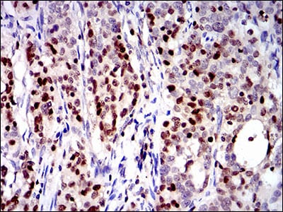
Figure 5: Immunohistochemical analysis of paraffin-embedded cervical cancer tissues using RUNX3 mouse mAb with DAB staining.
Elisa

Black line: Control Antigen (100 ng); Purple line: Antigen(10ng); Blue line: Antigen (50 ng); Red line: Antigen (100 ng);
For Research Use Only. Not for use in diagnostic procedures.