

RSK2 Primary Antibody
Item Information
Catalog #
Size
Price
Description
This gene encodes a member of the RSK (ribosomal S6 kinase) family of serine/threonine kinases. This kinase contains 2 non-identical kinase catalytic domains and phosphorylates various substrates, including members of the mitogen-activated kinase (MAPK) signalling pathway. The activity of this protein has been implicated in controlling cell growth and differentiation. Mutations in this gene have been associated with Coffin-Lowry syndrome (CLS).
Product Overview
Entrez GenelD
6197
Aliases
RPS6KA3; CLS; RSK; HU-3; RSK2; MRX19; ISPK-1; p90-RSK2; pp90RSK2; MAPKAPK1B; S6K-alpha3
Clone#
4E10
Host / Isotype
Mouse / IgG1
Species Reactivity
Human
Immunogen
Purified recombinant fragment of human RSK2 expressed in E. Coli.
Formulation
Ascitic fluid containing 0.03% sodium azide.
Storage
Store at 4°C short term. Aliquot and store at -20°C long term. Avoid freeze/thaw cycles.
Product Applications
WB (Western Blot)
1/500 - 1/2000
ICC (Immunocytochemistry)
1/200 - 1/1000
FCM (Flow Cytometry)
1/200 - 1/400
ELISA
1/10000
References
1. Mol Cell. 2009 Jan 16;33(1):109-16.
2. Cancer Res. 2009 May 15;69(10):4398-406.
2. Cancer Res. 2009 May 15;69(10):4398-406.
Product Image
Western Blot

Figure 1: Western blot analysis using RSK2 mAb against human RSK2 (AA: 1-212) recombinant protein. (Expected MW is 49.7 kDa)
Western Blot
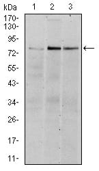
Figure 2: Western blot analysis using RSK2 mouse mAb against Hela (1), MCF-7 (2), and HepG2 (3) cell lysate.
Immunofluorescence analysis
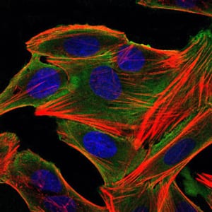
Figure 3: Immunofluorescence analysis of HepG2 cells using RSK2 mouse mAb (green). Blue: DRAQ5 fluorescent DNA dye. Red: Actin filaments have been labeled with Alexa Fluor-555 phalloidin.
Flow cytometric
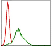
Figure 4: Flow cytometric analysis of HepG2 cells using RSK2 mouse mAb (green) and negative control (red).
Elisa

Black line: Control Antigen (100 ng); Purple line: Antigen(10ng); Blue line: Antigen (50 ng); Red line: Antigen (100 ng);
For Research Use Only. Not for use in diagnostic procedures.