

RPTOR Primary Antibody
Item Information
Catalog #
Size
Price
Description
This gene encodes a component of a signaling pathway that regulates cell growth in response to nutrient and insulin levels. The encoded protein forms a stoichiometric complex with the mTOR kinase, and also associates with eukaryotic initiation factor 4E-binding protein-1 and ribosomal protein S6 kinase. The protein positively regulates the downstream effector ribosomal protein S6 kinase, and negatively regulates the mTOR kinase. Multiple transcript variants encoding different isoforms have been found for this gene.
Product Overview
Entrez GenelD
57521
Aliases
KOG1; Mip1
Clone#
5A3A4
Host / Isotype
Mouse / IgG1
Species Reactivity
Human
Immunogen
Purified recombinant fragment of human RPTOR (AA: 874-1009) expressed in E. Coli.
Formulation
Purified antibody from tissue culture in PBS with 0.05% sodium azide
Storage
Store at 4°C short term. Aliquot and store at -20°C long term. Avoid freeze/thaw cycles.
Product Applications
WB (Western Blot)
1/500 - 1/2000
IHC_P(Immunohistochemistry)
1/200 - 1/1000
ICC (Immunocytochemistry)
1/200 - 1/1000
ELISA
1/10000
References
1. PLoS Genet. 2010 Oct 28;6(10):e1001178.
2. Cell Cycle. 2011 Sep 15;10(18):3140-52.
2. Cell Cycle. 2011 Sep 15;10(18):3140-52.
Product Image
Western Blot

Figure 1: Western blot analysis using RPTOR mAb against human RPTOR (AA: 874-1009) recombinant protein. (Expected MW is 41 kDa)
Western Blot
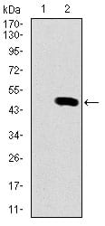
Figure 2: Western blot analysis using RPTOR mAb against HEK293 (1) and RPTOR (AA: 874-1009)-hIgGFc transfected HEK293 (2) cell lysate.
Immunofluorescence analysis
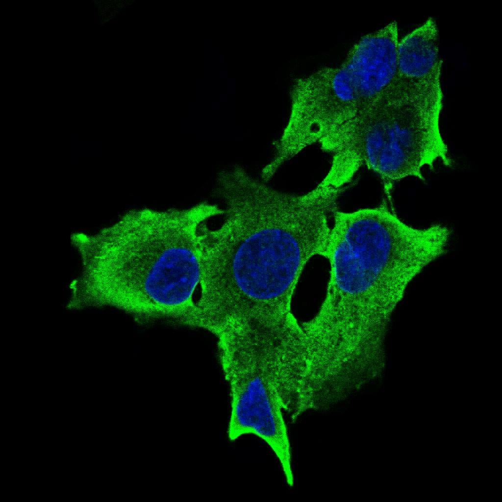
Figure 3: Immunofluorescence analysis of HepG2 cells using RPTOR mouse mAb (green). Blue: DRAQ5 fluorescent DNA dye. Secondary antibody from Fisher (Cat#: 35503)
Immunohistochemical analysis
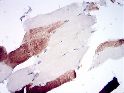
Figure 4: Immunohistochemical analysis of paraffin-embedded muscle tissues using RPTOR mouse mAb with DAB staining.
Elisa

Black line: Control Antigen (100 ng); Purple line: Antigen(10ng); Blue line: Antigen (50 ng); Red line: Antigen (100 ng);
For Research Use Only. Not for use in diagnostic procedures.