
RPS6KB1 Primary Antibody
Item Information
Catalog #
Size
Price
Description
This gene encodes a member of the RSK (ribosomal S6 kinase) family of serine/threonine kinases. This kinase contains 2 non-identical kinase catalytic domains and phosphorylates several residues of the S6 ribosomal protein. The kinase activity of this protein leads to an increase in protein synthesis and cell proliferation. Amplification of the region of DNA encoding this gene and overexpression of this kinase are seen in some breast cancer cell lines. Alternate translational start sites have been described and alternate transcriptional splice variants have been observed but have not been thoroughly characterized.
Product Overview
Entrez GenelD
6198
Aliases
S6K; PS6K; S6K1; STK14A; p70-S6K; p70-alpha; p70(S6K)-alpha
Clone#
5G9
Host / Isotype
Mouse / IgG1
Species Reactivity
Human
Immunogen
Purified recombinant fragment of human RPS6KB1 expressed in E. Coli.
Formulation
Purified antibody in PBS with 0.05% sodium azide
Storage
Store at 4°C short term. Aliquot and store at -20°C long term. Avoid freeze/thaw cycles.
Product Applications
WB (Western Blot)
1/500 - 1/2000
IHC_P(Immunohistochemistry)
1/200 - 1/1000
FCM (Flow Cytometry)
1/200 - 1/400
ELISA
1/10000
References
1. J Biol Chem. 2010 Jan 1;285(1):30-42.
2. Cell Mol Life Sci. 2009 Apr;66(8):1457-66.
2. Cell Mol Life Sci. 2009 Apr;66(8):1457-66.
Product Image
Western Blot

Figure 1: Western blot analysis using RPS6KB1 mAb against human RPS6KB1 (AA: 295-524) recombinant protein. (Expected MW is 59 kDa)
Immunohistochemical analysis
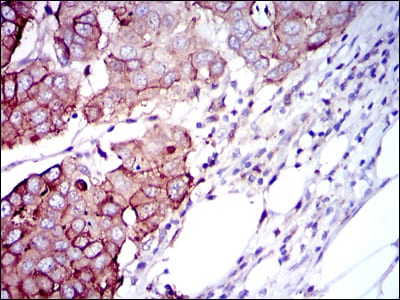
Figure 2: Immunohistochemical analysis of paraffin-embedded breast cancer tissues using RPS6KB1 mouse mAb with DAB staining.
Immunohistochemical analysis

Figure 3: Immunohistochemical analysis of paraffin-embedded muscle tissues using RPS6KB1 mouse mAb with DAB staining.
Flow cytometric
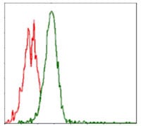
Figure 4: Flow cytometric analysis of Jurkat cells using RPS6KB1 mouse mAb (green) and negative control (red).
Elisa

Black line: Control Antigen (100 ng); Purple line: Antigen(10ng); Blue line: Antigen (50 ng); Red line: Antigen (100 ng);
For Research Use Only. Not for use in diagnostic procedures.