

RPS6KA2 Primary Antibody
Item Information
Catalog #
Size
Price
Description
This gene encodes a member of the RSK (ribosomal S6 kinase) family of serine/threonine kinases. This kinase contains 2 non-identical kinase catalytic domains and phosphorylates various substrates, including members of the mitogen-activated kinase (MAPK) signalling pathway. The activity of this protein has been implicated in controlling cell growth and differentiation. Alternate transcriptional splice variants, encoding different isoforms, have been characterized.
Product Overview
Entrez GenelD
6196
Aliases
RSK; HU-2; RSK3; p90-RSK3; pp90RSK3; MAPKAPK1C; S6K-alpha; S6K-alpha2
Clone#
3C4C8
Host / Isotype
Mouse / IgG1
Species Reactivity
Human, Mouse
Immunogen
Purified recombinant fragment of human RPS6KA2 (AA: 415-734) expressed in E. Coli.
Formulation
Purified antibody in PBS with 0.05% sodium azide.
Storage
Store at 4°C short term. Aliquot and store at -20°C long term. Avoid freeze/thaw cycles.
Product Applications
WB (Western Blot)
1/500 - 1/2000
IHC_P(Immunohistochemistry)
1/200 - 1/1000
ICC (Immunocytochemistry)
1/200 - 1/1000
FCM (Flow Cytometry)
1/200 - 1/400
ELISA
1/10000
References
1. Oncogene. 2007 Feb 1;26(5):683-700.
2. Exp Mol Med. 2003 Oct 31;35(5):365-70.
2. Exp Mol Med. 2003 Oct 31;35(5):365-70.
Product Image
Western Blot
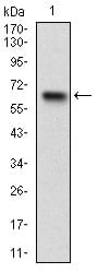
Figure 1: Western blot analysis using RPS6KA2 mAb against human RPS6KA2 (AA: 415-734) recombinant protein. (Expected MW is 62 kDa)
Western Blot

Figure 2: Western blot analysis using RPS6KA2 mAb against HEK293 (1) and RPS6KA2 (AA: 415-734)-hIgGFc transfected HEK293 (2) cell lysate.
Western Blot

Figure 3: Western blot analysis using RPS6KA2 mouse mAb against Hela (1), A431 (2), HEK293 (3), Jurkat (4), HepG2 (5), MCF-7 (6), NIH/3T3 (7) cell lysate.
Immunofluorescence analysis

Figure 4:Immunofluorescence analysis of A431 cells using RPS6KA2 mouse mAb (green). Blue: DRAQ5 fluorescent DNA dye. Red: Actin filaments have been labeled with Alexa Fluor- 555 phalloidin. Secondary antibody from Fisher (Cat#: 35503)
Flow cytometric

Figure 5: Flow cytometric analysis of Hela cells using RPS6KA2 mouse mAb (green) and negative control (red).
Immunohistochemical analysis

Figure 6: Immunohistochemical analysis of paraffin-embedded cervical cancer tissues using RPS6KA2 mouse mAb with DAB staining.
Immunohistochemical analysis

Figure 7: Immunohistochemical analysis of paraffin-embedded lung cancer tissues using RPS6KA2 mouse mAb with DAB staining.
Elisa

Black line: Control Antigen (100 ng); Purple line: Antigen(10ng); Blue line: Antigen (50 ng); Red line: Antigen (100 ng);
For Research Use Only. Not for use in diagnostic procedures.