
RND3 Primary Antibody
Item Information
Catalog #
Size
Price
Description
This gene encodes a protein which is a member of the small GTPase protein superfamily. The encoded protein binds only GTP but has no GTPase activity, and appears to act as a negative regulator of cytoskeletal organization leading to loss of adhesion. Multiple alternatively spliced variants, encoding the same protein, have been identified.
Product Overview
Entrez GenelD
390
Aliases
ARHE; Rho8; RhoE; memB
Clone#
5C7E8
Host / Isotype
Mouse / IgG1
Species Reactivity
Human
Immunogen
Purified recombinant fragment of human RND3 (AA: 104-241) expressed in E. Coli.
Formulation
Purified antibody in PBS with 0.05% sodium azide
Storage
Store at 4°C short term. Aliquot and store at -20°C long term. Avoid freeze/thaw cycles.
Product Applications
WB (Western Blot)
1/500 - 1/2000
IHC_P(Immunohistochemistry)
1/200 - 1/1000
ICC (Immunocytochemistry)
1/200 - 1/1000
FCM (Flow Cytometry)
1/200 - 1/400
ELISA
1/10000
References
1.Hepatology. 2013 Jan;57(1):152-61.
2.Oncol Rep. 2011 Jan;25(1):173-80.
2.Oncol Rep. 2011 Jan;25(1):173-80.
Product Image
Elisa

Figure 1: Black line: Control Antigen (100 ng); Purple line: Antigen(10ng); Blue line: Antigen (50 ng); Red line: Antigen (100 ng);
Western Blot
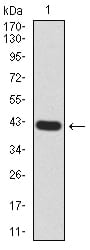
Figure 2:Western blot analysis using RND3 mAb against human RND3 (AA: 104-241) recombinant protein. (Expected MW is 41.3 kDa)
Western Blot

Figure 3:Western blot analysis using RND3 mAb against HEK293 (1) and RND3 (AA: 104-241)-hIgGFc transfected HEK293 (2) cell lysate.
Immunofluorescence analysis

Figure 4:Immunofluorescence analysis of Hela cells using RND3 mouse mAb (green). Blue: DRAQ5 fluorescent DNA dye. Red: Actin filaments have been labeled with Alexa Fluor- 555 phalloidin. Secondary antibody from Fisher (Cat#: 35503)
Flow cytometric

Figure 5:Flow cytometric analysis of Hela cells using RND3 mouse mAb (green) and negative control (red).
Immunohistochemical analysis

Figure 6:Immunohistochemical analysis of paraffin-embedded bladder cancer tissues using RND3 mouse mAb with DAB staining.
For Research Use Only. Not for use in diagnostic procedures.