

Ring1 Primary Antibody
Item Information
Catalog #
Size
Price
Description
This gene belongs to the RING finger family, members of which encode proteins characterized by a RING domain, a zinc-binding motif related to the zinc finger domain. The gene product can bind DNA and can act as a transcriptional repressor. It is associated with the multimeric polycomb group protein complex. The gene product interacts with the polycomb group proteins BMI1, EDR1, and CBX4, and colocalizes with these proteins in large nuclear domains. It interacts with the CBX4 protein via its glycine-rich C-terminal domain. The gene maps to the HLA class II region, where it is contiguous with the RING finger genes FABGL and HKE4.
Product Overview
Entrez GenelD
6015
Aliases
RNF1; RING1A
Clone#
8E8A2
Host / Isotype
Mouse / IgG1
Species Reactivity
Human
Immunogen
Purified recombinant fragment of human Ring1 (AA: 79-263) expressed in E. Coli.
Formulation
Purified antibody in PBS with 0.05% sodium azide
Storage
Store at 4°C short term. Aliquot and store at -20°C long term. Avoid freeze/thaw cycles.
Product Applications
WB (Western Blot)
1/500 - 1/2000
ICC (Immunocytochemistry)
1/200 - 1/1000
ELISA
1/10000
References
1. Int J Dev Biol. 2009;53(2-3):355-70.
2. PLoS One. 2009 Dec 1;4(12):e8104.
2. PLoS One. 2009 Dec 1;4(12):e8104.
Product Image
Western Blot

Figure 1: Western blot analysis using Ring1 mAb against human Ring1 recombinant protein. (Expected MW is 44.6 kDa)
Western Blot

Figure 2: Western blot analysis using Ring1 mAb against HEK293 (1) and Ring1 (AA: 79-263)-hIgGFc transfected HEK293 (2) cell lysate.
Immunofluorescence analysis

Figure 3: Immunofluorescence analysis of A431 cells using Ring1 mouse mAb (green). Blue: DRAQ5 fluorescent DNA dye. Red: Actin filaments have been labeled with Alexa Fluor-555 phalloidin.
Immunofluorescence analysis

Figure 3: Immunofluorescence analysis of Hela cells using Ring1 mouse mAb (green). Blue: DRAQ5 fluorescent DNA dye. Red: Actin filaments have been labeled with Alexa Fluor-555 phalloidin.
Elisa
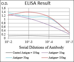
Black line: Control Antigen (100 ng); Purple line: Antigen(10ng); Blue line: Antigen (50 ng); Red line: Antigen (100 ng);
For Research Use Only. Not for use in diagnostic procedures.