
Mouse Monoclonal Antibody to RHOA
Item Information
Catalog #
Size
Price
Description
This gene encodes a member of the Rho family of small GTPases, which cycle between inactive GDP-bound and active GTP-bound states and function as molecular switches in signal transduction cascades. Rho proteins promote reorganization of the actin cytoskeleton and regulate cell shape, attachment, and motility. Overexpression of this gene is associated with tumor cell proliferation and metastasis. Multiple alternatively spliced variants have been identified. [provided by RefSeq, Sep 2015]
Product Overview
Entrez GenelD
387
Aliases
ARHA; ARH12; RHO12; EDFAOB; RHOH12
Clone#
7E7H4
Host / Isotype
Mouse / IgG2b
Immunogen
Purified recombinant fragment of human RHOA (AA: 1-190) expressed in E. Coli.
Formulation
Purified antibody in PBS with 0.05% sodium azide
Storage
Store at 4°C short term. Aliquot and store at -20°C long term. Avoid freeze/thaw cycles.
Product Applications
WB (Western Blot)
1/500 - 1/2000
IHC_P(Immunohistochemistry)
1/200 - 1/1000
ICC (Immunocytochemistry)
1/200 - 1/1000
FCM (Flow Cytometry)
1/200 - 1/400
ELISA
1/10000
References
1.Immunity.2020 Jul 14;53(1):187-203.e8.2.Nat Commun.2021 Apr 13;12(1):2226.
Product Image
Elisa

Figure 1:Black line: Control Antigen (100 ng);Purple line: Antigen (10ng); Blue line: Antigen (50 ng); Red line:Antigen (100 ng)
Western Blot

Figure 2:Western blot analysis using RHOA mAb against human RHOA (AA: 1-190) recombinant protein. (Expected MW is *** kDa)
Western Blot

Figure 3:Western blot analysis using RHOA mAb against HEK293 (1) and RHOA (AA: 1-190)-hIgGFc transfected HEK293-6e (2) cell lysate.
Western Blot

Figure 4:Western blot analysis using RHOA mouse mAb against Jurlat (1), MCF-7 (2),A431 (3), and Hela (4) cell lysate.
Immunofluorescence analysis

Figure 5:Immunofluorescence analysis of Hela cells using RHOA mouse mAb (green). Blue: DRAQ5 fluorescent DNA dye. Red: Actin filaments have been labeled with Alexa Fluor- 555 phalloidin. Secondary antibody from Fisher (Cat#: 35503)
Flow cytometric analysis

Figure 6:Flow cytometric analysis of Hela cells using RHOA mouse mAb (green) and negative control (red).
Flow cytometric analysis

Figure 7:Flow cytometric analysis of HepG2 cells using RHOA mouse mAb (green) and negative control (red).
Flow cytometric analysis

Figure 8:Flow cytometric analysis of Jurkat cells using RHOA mouse mAb (green) and negative control (red).
Flow cytometric analysis

Figure 9:Flow cytometric analysis of K562 cells using RHOA mouse mAb (green) and negative control (red).
Immunohistochemical analysis

Figure 10:Immunohistochemical analysis of paraffin-embedded gastric cancer tissues using RHOA mouse mAb with DAB staining.
Immunohistochemical analysis
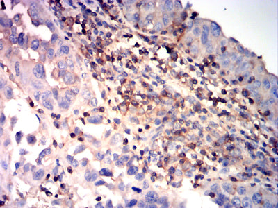
Figure 11:Immunohistochemical analysis of paraffin-embedded endometrial carcinoma tissues using RHOA mouse mAb with DAB staining.
For Research Use Only. Not for use in diagnostic procedures.