

RF1 Primary Antibody
Item Information
Catalog #
Size
Price
Description
This gene encodes a class-1 polypeptide chain release factor. The encoded protein plays an essential role in directing termination of mRNA translation from the termination codons UAA, UAG and UGA. This protein is a component of the SURF complex which promotes degradation of prematurely terminated mRNAs via the mechanism of nonsense-mediated mRNA decay (NMD). Alternate splicing results in multiple transcript variants. Pseudogenes of this gene are found on chromosomes 6, 7, and X.
Product Overview
Entrez GenelD
2107
Aliases
ERF; ETF1; ERF1; TB3-1; D5S1995; SUP45L1
Clone#
4F9H12
Host / Isotype
Mouse / IgG1
Species Reactivity
Human
Immunogen
Purified recombinant fragment of human RF1 (AA: 288-437) expressed in E. Coli.
Formulation
Purified antibody in PBS with 0.05% sodium azide
Storage
Store at 4°C short term. Aliquot and store at -20°C long term. Avoid freeze/thaw cycles.
Product Applications
WB (Western Blot)
1/500 - 1/2000
FCM (Flow Cytometry)
1/200 - 1/400
ELISA
1/10000
References
1.Nucleic Acids Res. 2013 Apr;41(8):4573-86.
2.Protein Sci. 2012 Jun;21(6):896-903.
2.Protein Sci. 2012 Jun;21(6):896-903.
Product Image
Elisa

Figure 1: Black line: Control Antigen (100 ng);Purple line: Antigen (10ng); Blue line: Antigen (50 ng); Red line:Antigen (100 ng)
Western Blot

Figure 2:Western blot analysis using RF1 mAb against human RF1 (AA: 288-437) recombinant protein. (Expected MW is 43 kDa)
Western Blot

Figure 3:Western blot analysis using RF1 mAb against HEK293 (1) and RF1 (AA: 288-437)-hIgGFc transfected HEK293 (2) cell lysate.
Flow cytometric

Figure 5:Flow cytometric analysis of Hela cells using RF1 mouse mAb (green) and negative control (red).
Flow cytometric
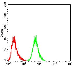
Figure 6:Flow cytometric analysis of HepG2 cells using RF1 mouse mAb (green) and negative control (red).
Western Blot

Figure 6:Western blot analysis using *** mouse mAb against MCF-7 (1), T47D (2), MOLT4 (3), and Raji (4) cell lysate.
For Research Use Only. Not for use in diagnostic procedures.