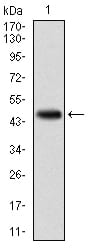
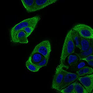
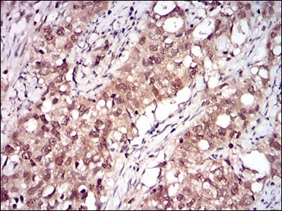
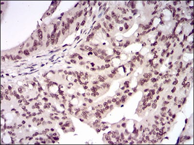
RELB Primary Antibody

Item Information
Catalog #
Size
Price
Description

NF-kappa-B is a pleiotropic transcription factor which is present in almost all cell types and is involved in many biological processed such as inflammation, immunity, differentiation, cell growth, tumorigenesis and apoptosis. NF-kappa-B is a homo- or heterodimeric complex formed by the Rel-like domain-containing proteins RELA/p65, RELB, NFKB1/p105, NFKB1/p50, REL and NFKB2/p52. The dimers bind at kappa-B sites in the DNA of their target genes and the individual dimers have distinct preferences for different kappa-B sites that they can bind with distinguishable affinity and specificity. Different dimer combinations act as transcriptional activators or repressors, respectively. NF-kappa-B is controlled by various mechanisms of post-translational modification and subcellular compartmentalization as well as by interactions with other cofactors or corepressors. NF-kappa-B complexes are held in the cytoplasm in an inactive state complexed with members of the NF-kappa-B inhibitor (I-kappa-B) family. In a conventional activation pathway, I-kappa-B is phosphorylated by I-kappa-B kinases (IKKs) in response to different activators, subsequently degraded thus liberating the active NF-kappa-B complex which translocates to the nucleus. NF-kappa-B heterodimeric RelB-p50 and RelB-p52 complexes are transcriptional activators. RELB neither associates with DNA nor with RELA/p65 or REL. Stimulates promoter activity in the presence of NFKB2/p49. As a member of the NUPR1/RELB/IER3 survival pathway, may provide pancreatic ductal adenocarcinoma with remarkable resistance to cell stress, such as starvation or gemcitabine treatment

Product Overview
Entrez GenelD
5971
Aliases
IREL; I-REL; REL-B
Clone#
2E4B1
Host / Isotype
Mouse / IgG2a
Species Reactivity
Human
Immunogen
Purified recombinant fragment of human RELB (AA: 273-443) expressed in E. Coli.
Formulation
Purified antibody from tissue culture in PBS with 0.05% sodium azide
Storage
4°C; -20°C for long term storage
Product Applications
WB (Western Blot)
1/500 - 1/2000
IHC_P(Immunohistochemistry)
1/200 - 1/1000
ICC (Immunocytochemistry)
1/200 - 1/1000
FCM (Flow Cytometry)
1/200 - 1/400
ELISA
1/10000
References
1. BMC Med Genomics. 2010 Nov 10;3:53.
2. Cancer Res. 2009 Apr 15;69(8):3267-71.
Product Image
Western Blot
Figure 1: Western blot analysis using RELB mAb against human RELB (AA: 273-443) recombinant protein. (Expected MW is 45.5 kDa)
Western Blot
Figure 2: Western blot analysis using RELB mAb against HEK293 (1) and RELB (AA: 273-443)-hIgGFc transfected HEK293 (2) cell lysate.
Immunofluorescence analysis
Figure 3: Immunofluorescence analysis of MCF-7 cells using RELB mouse mAb (green). Blue: DRAQ5 fluorescent DNA dye. Secondary antibody from Fisher (Cat#: 35503)
Flow cytometric
Figure 4: Flow cytometric analysis of MCF-7 cells using RELB mouse mAb (green) and negative control (red).
Immunohistochemical analysis
Figure 5: Immunohistochemical analysis of paraffin-embedded cervical cancer tissues using RELB mouse mAb with DAB staining.
Immunohistochemical analysis
Figure 6: Immunohistochemical analysis of paraffin-embedded rectum cancer tissues using RELB mouse mAb with DAB staining.
Elisa
Black line: Control Antigen (100 ng); Purple line: Antigen(10ng); Blue line: Antigen (50 ng); Red line: Antigen (100 ng);
For Research Use Only. Not for use in diagnostic procedures.