

RB1 Primary Antibody
Item Information
Catalog #
Size
Price
Description
The protein encoded by this gene is a negative regulator of the cell cycle and was the first tumor suppressor gene found. The encoded protein also stabilizes constitutive heterochromatin to maintain the overall chromatin structure. The active, hypophosphorylated form of the protein binds transcription factor E2F1. Defects in this gene are a cause of childhood cancer retinoblastoma (RB), bladder cancer, and osteogenic sarcoma.
Product Overview
Entrez GenelD
5925
Aliases
RB; pRb; OSRC; pp110; p105-Rb
Clone#
2A11D11
Host / Isotype
Mouse / IgG1
Species Reactivity
Human
Immunogen
Purified recombinant fragment of human RB1 (AA: 2106-2784) expressed in E. Coli.
Formulation
Purified antibody in PBS with 0.05% sodium azide
Storage
Store at 4°C short term. Aliquot and store at -20°C long term. Avoid freeze/thaw cycles.
Product Applications
WB (Western Blot)
1/500 - 1/2000
ICC (Immunocytochemistry)
1/200 - 1/1000
FCM (Flow Cytometry)
1/200 - 1/400
ELISA
1/10000
References
1. Cancer Genet. 2011 Jun;204(6):316-22.
2. J Neuropathol Exp Neurol. 2012 Jan;71(1):83-9.
2. J Neuropathol Exp Neurol. 2012 Jan;71(1):83-9.
Product Image
Western Blot

Figure 1: Western blot analysis using RB1 mAb against human RB1 (AA: 2106-2784) recombinant protein. (Expected MW is 38.8 kDa)
Western Blot
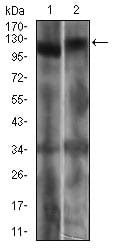
Figure 2: Western blot analysis using RB1 mouse mAb against Jurkat (1) and A431 (2) cell lysate.
Immunofluorescence analysis

Figure 3: Immunofluorescence analysis of NIH/3T3 cells using RB1 mouse mAb (green). Blue: DRAQ5 fluorescent DNA dye. Red: Actin filaments have been labeled with Alexa Fluor-555 phalloidin. Secondary antibody from Fisher (Cat#: 35503)
Flow cytometric

Figure 4: Flow cytometric analysis of Jurkat cells using RB1 mouse mAb (green) and negative control (red).
Elisa

Black line: Control Antigen (100 ng); Purple line: Antigen(10ng); Blue line: Antigen (50 ng); Red line: Antigen (100 ng);
For Research Use Only. Not for use in diagnostic procedures.