

RANBP9 Primary Antibody
Item Information
Catalog #
Size
Price
Description
This gene encodes a protein that binds RAN, a small GTP binding protein belonging to the RAS superfamily that is essential for the translocation of RNA and proteins through the nuclear pore complex. The protein encoded by this gene has also been shown to interact with several other proteins, including met proto-oncogene, homeodomain interacting protein kinase 2, androgen receptor, and cyclin-dependent kinase 11.
Product Overview
Entrez GenelD
10048
Aliases
BPM-L; BPM90; RANBPM; RanBP7
Clone#
6E10H5
Host / Isotype
Mouse / IgG1
Species Reactivity
Human, Mouse
Immunogen
Purified recombinant fragment of human RANBP9 (AA: 453-680) expressed in E. Coli.
Formulation
Purified antibody from tissue culture in PBS with 0.05% sodium azide
Storage
Store at 4°C short term. Aliquot and store at -20°C long term. Avoid freeze/thaw cycles.
Product Applications
WB (Western Blot)
1/500 - 1/2000
IHC_P(Immunohistochemistry)
1/200 - 1/1000
FCM (Flow Cytometry)
1/200 - 1/400
ELISA
1/10000
References
1. Biochem J. 2006 May 15;396(1):23-30.
2. BMC Cancer. 2002 Nov 6;2:28.
2. BMC Cancer. 2002 Nov 6;2:28.
Product Image
Western Blot
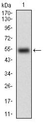
Figure 1: Western blot analysis using RANBP9 mAb against human RANBP9 (AA: 453-680) recombinant protein. (Expected MW is 50.9 kDa)
Western Blot
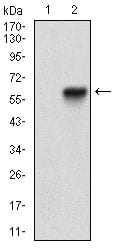
Figure 2: Western blot analysis using RANBP9 mAb against HEK293 (1) and RANBP9 (AA: 453-680)-hIgGFc transfected HEK293 (2) cell lysate.
Western Blot
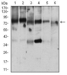
Figure 3: Western blot analysis using RANBP9 mouse mAb against Jurkat (1), MOLT4 (2), HEK293 (3), A431 (4), A549 (5), NIH/3T3 (6) cell lysate.
Flow cytometric
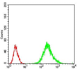
Figure 4: Flow cytometric analysis of Jurkat cells using RANBP9 mouse mAb (green) and negative control (red).
Immunohistochemical analysis
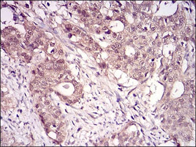
Figure 5: Immunohistochemical analysis of paraffin-embedded cervical cancer tissues using RANBP9 mouse mAb with DAB staining.
Immunohistochemical analysis
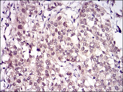
Figure 6: Immunohistochemical analysis of paraffin-embedded bladder cancer tissues using RANBP9 mouse mAb with DAB staining.
Elisa
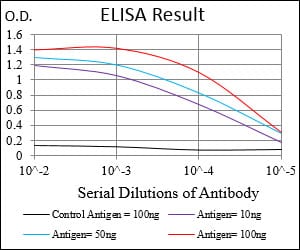
Black line: Control Antigen (100 ng); Purple line: Antigen(10ng); Blue line: Antigen (50 ng); Red line: Antigen (100 ng);
For Research Use Only. Not for use in diagnostic procedures.