

RAF1 Primary Antibody
Item Information
Catalog #
Size
Price
Description
This gene is the cellular homolog of viral raf gene (v-raf). The encoded protein is a MAP kinase kinase kinase (MAP3K), which functions downstream of the Ras family of membrane associated GTPases to which it binds directly. Once activated, the cellular RAF1 protein can phosphorylate to activate the dual specificity protein kinases MEK1 and MEK2, which in turn phosphorylate to activate the serine/threonine specific protein kinases, ERK1 and ERK2. Activated ERKs are pleiotropic effectors of cell physiology and play an important role in the control of gene expression involved in the cell division cycle, apoptosis, cell differentiation and cell migration. Mutations in this gene are associated with Noonan syndrome 5 and LEOPARD syndrome 2.
Product Overview
Entrez GenelD
5894
Aliases
NS5; CRAF; Raf-1; c-Raf
Clone#
4G4
Host / Isotype
Mouse / IgG1
Species Reactivity
Human
Immunogen
Purified recombinant fragment of human RAF1 expressed in E. Coli.
Formulation
Purified antibody in PBS with 0.05% sodium azide
Storage
Store at 4°C short term. Aliquot and store at -20°C long term. Avoid freeze/thaw cycles.
Product Applications
WB (Western Blot)
1/500 - 1/2000
IHC_P(Immunohistochemistry)
1/200 - 1/1000
FCM (Flow Cytometry)
1/200 - 1/400
ELISA
1/10000
References
1. Am J Hum Genet. 2009 Nov;85(5):628-42.
2. Mol Cancer Res. 2009 Oct;7(10):1635-44.
2. Mol Cancer Res. 2009 Oct;7(10):1635-44.
Product Image
Western Blot

Figure 1: Western blot analysis using RAF1 mAb against human RAF1 (AA: 198-407) recombinant protein. (Expected MW is 73 kDa)
Western Blot

Figure 2: Western blot analysis using RAF1 mouse mAb against Hela (1), A431 (2), Cos7 (3) and C6 (4) cell lysate.
Immunohistochemical analysis

Figure 3: Immunohistochemical analysis of paraffin-embedded liver cancer tissues using RAF1 mouse mAb with DAB staining.
Flow cytometric
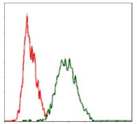
Figure 4: Flow cytometric analysis of Hela cells using RAF1 mouse mAb (green) and negative control (red).
Elisa

Black line: Control Antigen (100 ng); Purple line: Antigen(10ng); Blue line: Antigen (50 ng); Red line: Antigen (100 ng);
For Research Use Only. Not for use in diagnostic procedures.