

RAD50 Primary Antibody
Item Information
Catalog #
Size
Price
Description
The protein encoded by this gene is highly similar to Saccharomyces cerevisiae Rad50, a protein involved in DNA double-strand break repair. This protein forms a complex with MRE11 and NBS1. The protein complex binds to DNA and displays numerous enzymatic activities that are required for nonhomologous joining of DNA ends. This protein, cooperating with its partners, is important for DNA double-strand break repair, cell cycle checkpoint activation, telomere maintenance, and meiotic recombination. Knockout studies of the mouse homolog suggest this gene is essential for cell growth and viability. Mutations in this gene are the cause of Nijmegen breakage syndrome-like disorder.
Product Overview
Entrez GenelD
10111
Aliases
NBSLD; RAD502; hRad50
Clone#
3D2G10
Host / Isotype
Mouse / IgG2a
Species Reactivity
Human, Rat
Immunogen
Purified recombinant fragment of human RAD50 (AA: 228-359) expressed in E. Coli.
Formulation
Purified antibody in PBS with 0.05% sodium azide
Storage
Store at 4°C short term. Aliquot and store at -20°C long term. Avoid freeze/thaw cycles.
Product Applications
WB (Western Blot)
1/500 - 1/2000
IHC_P(Immunohistochemistry)
1/200 - 1/1000
FCM (Flow Cytometry)
1/200 - 1/400
ELISA
1/10000
References
1.Breast Cancer Res Treat. 2010 Sep;123(2):607-9.
2.Dis Markers. 2008;24(2):127-34.
2.Dis Markers. 2008;24(2):127-34.
Product Image
Elisa
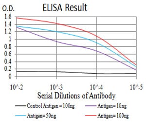
Figure 1: Black line: Control Antigen (100 ng);Purple line: Antigen (10ng); Blue line: Antigen (50 ng); Red line:Antigen (100 ng)
Western Blot
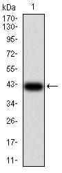
Figure 2:Western blot analysis using RAD50 mAb against human RAD50 (AA: 228-359) recombinant protein. (Expected MW is 41.7 kDa)
Western Blot
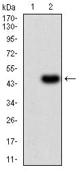
Figure 3:Western blot analysis using RAD50 mAb against HEK293 (1) and RAD50 (AA: 228-359)-hIgGFc transfected HEK293 (2) cell lysate.
Western Blot
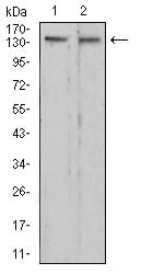
Figure 4:Western blot analysis using RAD50 mouse mAb against C6 (1) and HepG2 (2) cell lysate.
Flow cytometric
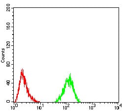
Figure 5:Flow cytometric analysis of Hela cells using RAD50 mouse mAb (green) and negative control (red).
Immunohistochemical analysis
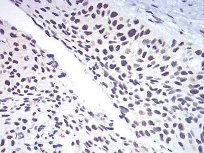
Figure 6:Immunohistochemical analysis of paraffin-embedded esophageal cancer tissues using RAD50 mouse mAb with DAB staining.
Immunohistochemical analysis
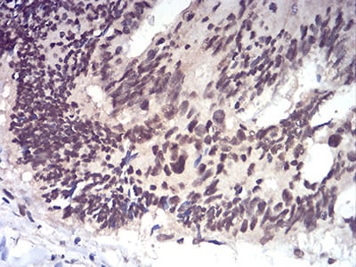
Figure 7:Immunohistochemical analysis of paraffin-embedded rectum cancer tissues using RAD50 mouse mAb with DAB staining.
For Research Use Only. Not for use in diagnostic procedures.