

RAD21 Primary Antibody
Item Information
Catalog #
Size
Price
Description
The protein encoded by this gene is highly similar to the gene product of Schizosaccharomyces pombe rad21, a gene involved in the repair of DNA double-strand breaks, as well as in chromatid cohesion during mitosis. This protein is a nuclear phospho-protein, which becomes hyperphosphorylated in cell cycle M phase. The highly regulated association of this protein with mitotic chromatin specifically at the centromere region suggests its role in sister chromatid cohesion in mitotic cells.
Product Overview
Entrez GenelD
5885
Aliases
HR21; MCD1; NXP1; SCC1; CDLS4; hHR21; HRAD21
Clone#
1B6D1
Host / Isotype
Mouse / IgG1
Species Reactivity
Human, Monkey, Rat
Immunogen
Purified recombinant fragment of human RAD21 (AA: 287-403) expressed in E. Coli.
Formulation
Purified antibody in PBS with 0.05% sodium azide
Storage
Store at 4°C short term. Aliquot and store at -20°C long term. Avoid freeze/thaw cycles.
Product Applications
WB (Western Blot)
1/500 - 1/2000
IHC_P(Immunohistochemistry)
1/200 - 1/1000
ICC (Immunocytochemistry)
1/200 - 1/1000
FCM (Flow Cytometry)
1/200 - 1/400
ELISA
1/10000
References
1.Breast Cancer Res. 2012 Apr 26;14(2):R69.
2.Breast Cancer Res. 2011 Jan 21;13(1):R9.
2.Breast Cancer Res. 2011 Jan 21;13(1):R9.
Product Image
Elisa
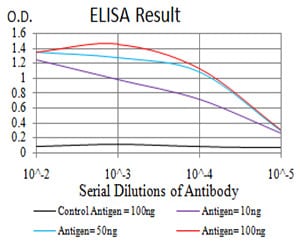
Figure 1: Black line: Control Antigen (100 ng);Purple line: Antigen (10ng); Blue line: Antigen (50 ng); Red line:Antigen (100 ng)
Western Blot
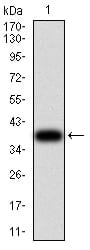
Figure 2:Western blot analysis using RAD21 mAb against human RAD21 (AA: 287-403) recombinant protein. (Expected MW is 39.3 kDa)
Western Blot
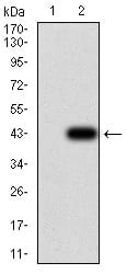
Figure 3:Western blot analysis using RAD21 mAb against HEK293 (1) and RAD21 (AA: 287-403)-hIgGFc transfected HEK293 (2) cell lysate.
Western Blot
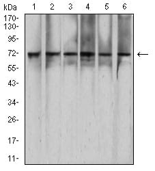
Figure 4:Western blot analysis using RAD21 mouse mAb against Hela (1), HEK293 (2), K562 (3), C6 (4), *** (5), and COS7 (6) cell lysate.
Immunofluorescence analysis
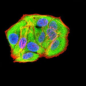
Figure 5:Immunofluorescence analysis of Hela cells using RAD21 mouse mAb (green). Blue: DRAQ5 fluorescent DNA dye. Red: Actin filaments have been labeled with Alexa Fluor- 555 phalloidin. Secondary antibody from Fisher (Cat#: 35503)
Flow cytometric
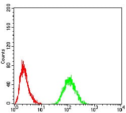
Figure 6:Flow cytometric analysis of Hela cells using RAD21 mouse mAb (green) and negative control (red).
Immunohistochemical analysis
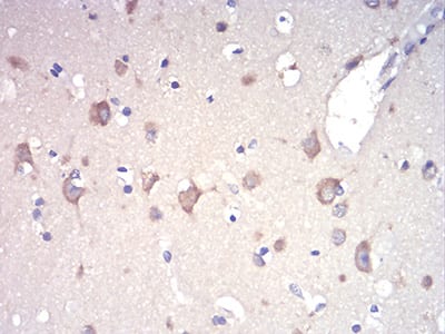
Figure 7:Immunohistochemical analysis of paraffin-embedded brain tissues using RAD21 mouse mAb with DAB staining.
Immunohistochemical analysis
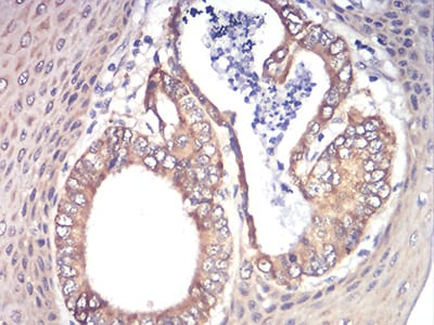
Figure 8:Immunohistochemical analysis of paraffin-embedded esophageal cancer tissues using RAD21 mouse mAb with DAB staining.
For Research Use Only. Not for use in diagnostic procedures.