
Rab5a Primary Antibody
Item Information
Catalog #
Size
Price
Description
RAB5A (RAB5A, Member RAS Oncogene Family) is a Protein Coding gene. Diseases associated with RAB5A include borna disease and choroideremia. Among its related pathways are Ras signaling pathway and Endocytosis. GO annotations related to this gene include GTP binding and GDP binding. An important paralog of this gene is RAB5C.
Product Overview
Entrez GenelD
5868
Aliases
RAB5
Clone#
3H5C11
Host / Isotype
Mouse / IgG1
Species Reactivity
Human
Immunogen
Purified recombinant fragment of human Rab5a (AA: 1-215) expressed in E. Coli.
Formulation
Purified antibody in PBS with 0.05% sodium azide
Storage
Store at 4°C short term. Aliquot and store at -20°C long term. Avoid freeze/thaw cycles.
Product Applications
WB (Western Blot)
1/500 - 1/2000
IHC_P(Immunohistochemistry)
1/200 - 1/1000
ICC (Immunocytochemistry)
1/200 - 1/1000
FCM (Flow Cytometry)
1/200 - 1/400
ELISA
1/10000
References
1.Cancer Sci. 2011 Dec;102(12):2172-8.
2.Cell Mol Life Sci. 2011 Aug;68(16):2785-95.
2.Cell Mol Life Sci. 2011 Aug;68(16):2785-95.
Product Image
Elisa

Figure 1: Black line: Control Antigen (100 ng); Purple line: Antigen(10ng); Blue line: Antigen (50 ng); Red line: Antigen (100 ng);
Western Blot
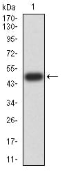
Figure 2:Western blot analysis using Rab5a mAb against human Rab5a (AA: 1-215) recombinant protein. (Expected MW is 49.5 kDa)
Western Blot

Figure 3:Western blot analysis using Rab5a mAb against HEK293 (1) and Rab5a (AA: 1-215)-hIgGFc transfected HEK293 (2) cell lysate.
Western Blot

Figure 4:Western blot analysis using Rab5a mouse mAb against Hela (1) and K562 (2) cell lysate.
Immunofluorescence analysis

Figure 5:Immunofluorescence analysis of Hela cells using Rab5a mouse mAb (green). Blue: DRAQ5 fluorescent DNA dye. Red: Actin filaments have been labeled with Alexa Fluor- 555 phalloidin. Secondary antibody from Fisher (Cat#: 35503)
Flow cytometric

Figure 6:Flow cytometric analysis of Hela cells using Rab5a mouse mAb (green) and negative control (red).
Immunohistochemical analysis
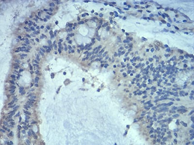
Figure 7:Immunohistochemical analysis of paraffin-embedded colon cancer tissues using Rab5a mouse mAb with DAB staining.
Immunohistochemical analysis

Figure 8:Immunohistochemical analysis of paraffin-embedded duodenum tissues using Rab5a mouse mAb with DAB staining.
For Research Use Only. Not for use in diagnostic procedures.