Rab25 Primary Antibody

Item Information
Catalog #
Size
Price
Description

Members of the Ras-related superfamily of GTP binding proteins, which includes Ras, Rho, Rab and ARF subfamilies, exhibit 30-50% similarity with Ras p21. Rab proteins play an important role for either in endocytosis or in biosynthetic protein transport. The possibility that Rab proteins might also direct the exocytosis from secretory vesicles to the plasma membrane is supported by the observation that in yeast, the SEC4 protein, which is 40% similar to Rab proteins, is associated with secretory vesicles. Rab proteins located on the cytoplasmic face of organelles and vesicles, rab proteins are involved in intracellular membrane fusion reactions. Rab25 was cloned from a gastric parietal cell cDNA library and is expressed in epithelial tissues such as the gastrointestinal mucosae, kidney, and lung, which encoded a protein of 28 kDa.

Product Overview
Entrez GenelD
57111
Aliases
CATX-8; RAB25
Clone#
3F12
Host / Isotype
Mouse / IgG1
Species Reactivity
Human, Mouse
Immunogen
Purified recombinant fragment of Rab25 expressed in E. Coli.
Formulation
Ascitic fluid containing 0.03% sodium azide.
Storage
4°C; -20°C for long term storage
Product Applications
WB (Western Blot)
1/500 - 1/2000
IHC_P(Immunohistochemistry)
1/200 - 1/1000
ICC (Immunocytochemistry)
1/200 - 1/1000
FCM (Flow Cytometry)
1/200 - 1/400
ELISA
1/10000
References
1. JR Goldenring, KR Shen, HD Vaughan. et al. J. Biol. Chem,1993,268(25):18419-18422
2. Xiaoye W, Ravindra K, Jennifer N. et al. J. Biol. Chem,2000,275(37):29138-29146
Product Image
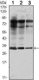
Western Blot
Figure 1: Western blot analysis using Rab25 mouse mAb against MCF-7 (1), T47D (2) and GC7901 (3) cell lysate.
Immunohistochemical analysis
Figure 2: Immunohistochemical analysis of paraffin-embedded esophagus tissues (left) and human lung cancer (right) using Rab25 mouse mAb with DAB staining.
Flow cytometric
Figure 3: Flow cytometric analysis of NIH/3T3 cells using Rab25 mouse mAb (green) and negative control (purple).
Immunofluorescence analysis
Figure 4: Immunofluorescence analysis of A549 cells using RAB25 mouse mAb (green). Blue: DRAQ5 fluorescent DNA dye. Red: Actin filaments have been labeled with Alexa Fluor-555 phalloidin.
For Research Use Only. Not for use in diagnostic procedures.