

Rab1b Primary Antibody
Item Information
Catalog #
Size
Price
Description
Members of the RAB protein family, such as RAB1B, are low molecular mass monomeric GTPases localized on the cytoplasmic surfaces of distinct membrane-bound organelles. RAB1B functions in the early secretory pathway and is essential for vesicle transport between the endoplasmic reticulum (ER) and Golgi
Product Overview
Entrez GenelD
81876
Aliases
N
Clone#
7A12G2
Host / Isotype
Mouse / IgG2b
Species Reactivity
Human
Immunogen
Purified recombinant fragment of human Rab1b (AA: 60-201) expressed in E. Coli.
Formulation
Purified antibody in PBS with 0.05% sodium azide
Storage
Store at 4°C short term. Aliquot and store at -20°C long term. Avoid freeze/thaw cycles.
Product Applications
WB (Western Blot)
1/500 - 1/2000
IHC_P(Immunohistochemistry)
1/200 - 1/1000
FCM (Flow Cytometry)
1/200 - 1/400
ELISA
1/10000
References
1.Mol Biol Cell. 2013 Mar;24(5):617-32.
2.Eur J Cell Biol. 2011 Apr;90(4):301-11.
2.Eur J Cell Biol. 2011 Apr;90(4):301-11.
Product Image
Elisa
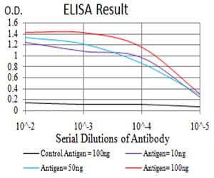
Figure 1: Black line: Control Antigen (100 ng);Purple line: Antigen (10ng); Blue line: Antigen (50 ng); Red line:Antigen (100 ng)
Western Blot
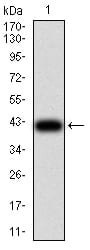
Figure 2:Western blot analysis using Rab1b mAb against human Rab1b (AA: 60-201) recombinant protein. (Expected MW is 41.4 kDa)
Western Blot
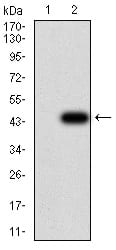
Figure 3:Western blot analysis using Rab1b mAb against HEK293 (1) and Rab1b (AA: 60-201)-hIgGFc transfected HEK293 (2) cell lysate.
Flow cytometric
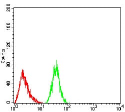
Figure 4:Flow cytometric analysis of Hela cells using Rab1b mouse mAb (green) and negative control (red).
Immunohistochemical analysis
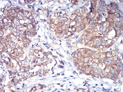
Figure 5:Immunohistochemical analysis of paraffin-embedded bladder cancer tissues using Rab1b mouse mAb with DAB staining.
Immunohistochemical analysis
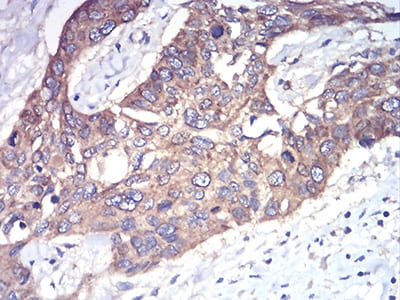
Figure 6:Immunohistochemical analysis of paraffin-embedded esophageal cancer tissues using Rab1b mouse mAb with DAB staining.
For Research Use Only. Not for use in diagnostic procedures.