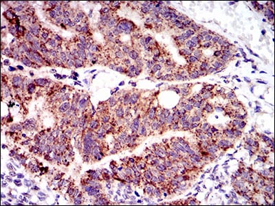
PTPN11 Primary Antibody

Item Information
Catalog #
Size
Price
Description

The protein encoded by this gene is a member of the protein tyrosine phosphatase (PTP) family. PTPs are known to be signaling molecules that regulate a variety of cellular processes including cell growth, differentiation, mitotic cycle, and oncogenic transformation. This PTP contains two tandem Src homology-2 domains, which function as phospho-tyrosine binding domains and mediate the interaction of this PTP with its substrates. This PTP is widely expressed in most tissues and plays a regulatory role in various cell signaling events that are important for a diversity of cell functions, such as mitogenic activation, metabolic control, transcription regulation, and cell migration. Mutations in this gene are a cause of Noonan syndrome as well as acute myeloid leukemia. Two transcript variants encoding different isoforms have been found for this gene.

Product Overview
Entrez GenelD
5781
Aliases
CFC; NS1; SHP2; BPTP3; PTP2C; PTP-1D; SH-PTP2; SH-PTP3; MGC14433
Clone#
6D9
Host / Isotype
Mouse / IgG1
Species Reactivity
Human
Immunogen
Purified recombinant fragment of human PTPN11 (AA: 263-329) expressed in E. Coli.
Formulation
Purified antibody in PBS with 0.05% sodium azide
Storage
4°C; -20°C for long term storage
Product Applications
WB (Western Blot)
1/500 - 1/2000
IHC_P(Immunohistochemistry)
1/200 - 1/1000
ICC (Immunocytochemistry)
1/200 - 1/1000
FCM (Flow Cytometry)
1/200 - 1/400
ELISA
1/10000
References
1.Blood. 2011 Aug 11;118(6):1504-15. 2.Cancer Cell. 2011 May 17;19(5):629-39.
Product Image
Western Blot
Figure 1: Western blot analysis using PTPN11 mAb against human PTPN11 recombinant protein. (Expected MW is 33.4 kDa)
Immunofluorescence analysis
Figure 2: Immunofluorescence analysis of HeLa cells using PTPN11 mouse mAb (green). Blue: DRAQ5 fluorescent DNA dye.
Flow cytometric
Figure 3: Flow cytometric analysis of HepG2 cells using PTPN11 mouse mAb (green) and negative control (red).
Immunohistochemical analysis
Figure 4: Immunohistochemical analysis of paraffin-embedded breast cancer tissues using PTPN11 mouse mAb with DAB staining.
Immunohistochemical analysis
Figure 5: Immunohistochemical analysis of paraffin-embedded rectum cancer tissues using PTPN11 mouse mAb with DAB staining.
Elisa
Black line: Control Antigen (100 ng); Purple line: Antigen(10ng); Blue line: Antigen (50 ng); Red line: Antigen (100 ng);
For Research Use Only. Not for use in diagnostic procedures.