
PSMC3 Primary Antibody
Item Information
Catalog #
Size
Price
Description
The 26S proteasome is a multicatalytic proteinase complex with a highly ordered structure composed of 2 complexes, a 20S core and a 19S regulator. The 20S core is composed of 4 rings of 28 non-identical subunits; 2 rings are composed of 7 alpha subunits and 2 rings are composed of 7 beta subunits. The 19S regulator is composed of a base, which contains 6 ATPase subunits and 2 non-ATPase subunits, and a lid, which contains up to 10 non-ATPase subunits. Proteasomes are distributed throughout eukaryotic cells at a high concentration and cleave peptides in an ATP/ubiquitin-dependent process in a non-lysosomal pathway. An essential function of a modified proteasome, the immunoproteasome, is the processing of class I MHC peptides. This gene encodes one of the ATPase subunits, a member of the triple-A family of ATPases that have chaperone-like activity. This subunit may compete with PSMC2 for binding to the HIV tat protein to regulate the interaction between the viral protein and the transcription complex. A pseudogene has been identified on chromosome 9.
Product Overview
Entrez GenelD
5702
Aliases
TBP1
Clone#
1G10C9
Host / Isotype
Mouse / IgG1
Species Reactivity
Human, Monkey, Rat
Immunogen
Purified recombinant fragment of human *** (AA: 53-152) expressed in E. Coli.
Formulation
Purified antibody in PBS with 0.05% sodium azide
Storage
Store at 4°C short term. Aliquot and store at -20°C long term. Avoid freeze/thaw cycles.
Product Applications
WB (Western Blot)
1/500 - 1/2000
IHC_P(Immunohistochemistry)
1/200 - 1/1000
ICC (Immunocytochemistry)
1/200 - 1/1000
FCM (Flow Cytometry)
1/200 - 1/400
ELISA
1/10000
References
1.Oncogene. 2012 Apr 5;31(14):1817-24.
2.PLoS One. 2011;6(10):e22800.
2.PLoS One. 2011;6(10):e22800.
Product Image
Elisa
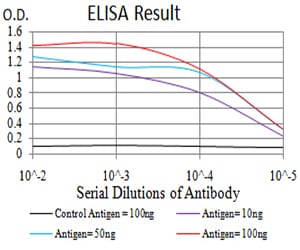
Figure 1: Black line: Control Antigen (100 ng);Purple line: Antigen (10ng); Blue line: Antigen (50 ng); Red line:Antigen (100 ng)
Western Blot

Figure 2:Western blot analysis using PSMC3 mAb against human PSMC3 (AA: 53-152) recombinant protein. (Expected MW is 37.2 kDa)
Western Blot
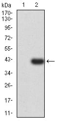
Figure 3:Western blot analysis using PSMC3 mAb against HEK293 (1) and PSMC3 (AA: 53-152)-hIgGFc transfected HEK293 (2) cell lysate.
Western Blot
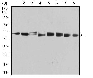
Figure 4:Western blot analysis using PSMC3 mouse mAb against MCF-7 (1), PC-3 (2), T47D (3), SW620 (4), COS7 (5), C6 (6), HELA (7), and A431 (8) cell lysate.
Immunofluorescence analysis
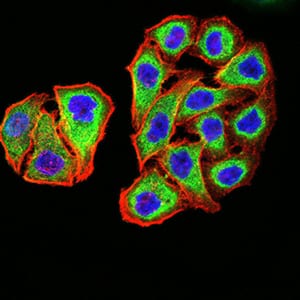
Figure 5:Immunofluorescence analysis of MCF-7 cells using PSMC3 mouse mAb (green). Blue: DRAQ5 fluorescent DNA dye. Red: Actin filaments have been labeled with Alexa Fluor- 555 phalloidin. Secondary antibody from Fisher (Cat#: 35503)
Immunofluorescence analysis
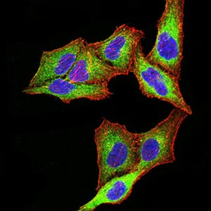
Figure 6:Immunofluorescence analysis of SK-OV-3 cells using PSMC3 mouse mAb (green). Blue: DRAQ5 fluorescent DNA dye. Red: Actin filaments have been labeled with Alexa Fluor- 555 phalloidin. Secondary antibody from Fisher (Cat#: 35503)
Flow cytometric
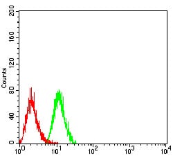
Figure 7:Flow cytometric analysis of Hela cells using PSMC3 mouse mAb (green) and negative control (red).
Immunohistochemical analysis
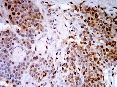
Figure 8:Immunohistochemical analysis of paraffin-embedded bladder cancer tissues using PSMC3 mouse mAb with DAB staining.
Immunohistochemical analysis
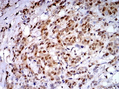
Figure 9:Immunohistochemical analysis of paraffin-embedded breast cancer tissues using PSMC3 mouse mAb with DAB staining.
For Research Use Only. Not for use in diagnostic procedures.