
CD202B Primary Antibody
Item Information
Catalog #
Size
Price
Description
This gene encodes a receptor that belongs to the protein tyrosine kinase Tie2 family. The encoded protein possesses a unique extracellular region that contains two immunoglobulin-like domains, three epidermal growth factor (EGF)-like domains and three fibronectin type III repeats. The ligand angiopoietin-1 binds to this receptor and mediates a signaling pathway that functions in embryonic vascular development. Mutations in this gene are associated with inherited venous malformations of the skin and mucous membranes. Alternative splicing results in multiple transcript variants. Additional alternatively spliced transcript variants of this gene have been described, but their full-length nature is not known.
Product Overview
Entrez GenelD
7010
Aliases
TEK; TIE2; VMCM; GLC3E; TIE-2; VMCM1
Clone#
3F5B9
Host / Isotype
Mouse / IgG2b
Species Reactivity
Human
Immunogen
Purified recombinant fragment of human CD202B (AA: extra 571-748) expressed in E. Coli.
Formulation
Purified antibody in PBS with 0.05% sodium azide
Storage
Store at 4°C short term. Aliquot and store at -20°C long term. Avoid freeze/thaw cycles.
Product Applications
WB (Western Blot)
1/500 - 1/2000
FCM (Flow Cytometry)
1/200 - 1/400
ELISA
1/10000
References
1.Oncotarget. 2016 Jan 19;7(3):2572-84.
2.Clin Res Cardiol. 2016 Aug;105(8):666-76.
2.Clin Res Cardiol. 2016 Aug;105(8):666-76.
Product Image
Elisa

Figure 1:Black line: Control Antigen (100 ng);Purple line: Antigen (10ng); Blue line: Antigen (50 ng); Red line:Antigen (100 ng)
Western Blot

Figure 2:Western blot analysis using CD202B mAb against human CD202B (AA: extra 571-748) recombinant protein. (Expected MW is 45.9 kDa)
Western Blot

Figure 3:Western blot analysis using CD202B mAb against HEK293 (1) and CD202B (AA: 23-745)-hIgGFc transfected HEK293 (2) cell lysate.
Flow cytometric
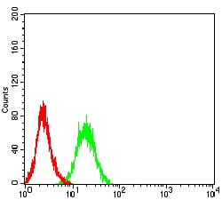
Figure 4:Flow cytometric analysis of HL-60 cells using CD202B mouse mAb (green) and negative control (red).
For Research Use Only. Not for use in diagnostic procedures.