PSA (KLK3) Primary Antibody

Item Information
Catalog #
Size
Price
Description

Kallikrein-related peptidase 3.Kallikreins are a subgroup of serine proteases having diverse physiological functions. Growing evidence suggests that many kallikreins are implicated in carcinogenesis and some have potential as novel cancer and other disease biomarkers. This gene is one of the fifteen kallikrein subfamily members located in a cluster on chromosome 19. Its protein product is a protease present in seminal plasma. It is thought to function normally in the liquefaction of seminal coagulum, presumably by hydrolysis of the high molecular mass seminal vesicle protein. Serum level of this protein, called PSA in the clinical setting, is useful in the diagnosis and monitoring of prostatic carcinoma. Alternate splicing of this gene generates several transcript variants encoding different isoforms.

Product Overview
Entrez GenelD
354
Aliases
APS; PSA; hK3; KLK2A1; KLK3
Clone#
5A11E2
Host / Isotype
Mouse / IgG1
Species Reactivity
Human
Immunogen
Purified recombinant fragment of KLK3 (aa26-251) expressed in E. Coli.
Formulation
Ascitic fluid containing 0.03% sodium azide.
Storage
4°C; -20°C for long term storage
Product Applications
WB (Western Blot)
1/500 - 1/2000
IHC_P(Immunohistochemistry)
1/200 - 1/1000
FCM (Flow Cytometry)
1/200 - 1/400
ELISA
1/10000
References
1. Am J Surg Pathol. 2007 Sep;31(9):1351-5.
2. Chem Biol Drug Des. 2007 Sep;70(3):261-7.
Product Image
Western Blot
Figure 1: Western blot analysis using KLK3 mouse mAb against truncated KLK3 recombinant protein.
Immunohistochemical analysis
Figure 2: Immunohistochemical analysis of paraffin-embedded human prostate carcinoma tissues, showing cytoplasmic localization using KLK3 mouse mAb with DAB staining.
Immunohistochemical analysis
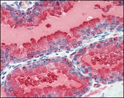
Figure 3: Immunohistochemical analysis of paraffin-embedded human prostate tissues using KLK3 mouse mAb with DAB staining.
Flow cytometric
Figure 4: Flow cytometric analysis of PC-3 cells using KLK3 mouse mAb (right) and negative control (left).
For Research Use Only. Not for use in diagnostic procedures.