
PRKDC Primary Antibody
Item Information
Catalog #
Size
Price
Description
The PRKDC gene encodes the catalytic subunit of a nuclear DNA-dependent serine/threonine protein kinase (DNA-PK). The second component is the autoimmune antigen Ku (MIM 152690), which is encoded by the G22P1 gene on chromosome 22q. On its own, the catalytic subunit of DNA-PK is inactive and relies on the G22P1 component to direct it to the DNA and trigger its kinase activity; PRKDC must be bound to DNA to express its catalytic properties.
Product Overview
Entrez GenelD
5591
Aliases
HYRC; p350; DNAPK; DNPK1; HYRC1; XRCC7; DNA-PKcs; PRKDC
Clone#
3H6
Host / Isotype
Mouse / IgG1
Species Reactivity
Human
Immunogen
Purified recombinant fragment of human PRKDC expressed in E. Coli.
Formulation
Ascitic fluid containing 0.03% sodium azide.
Storage
Store at 4°C short term. Aliquot and store at -20°C long term. Avoid freeze/thaw cycles.
Product Applications
WB (Western Blot)
1/500 - 1/2000
IHC_P(Immunohistochemistry)
1/200 - 1/1000
ICC (Immunocytochemistry)
1/200 - 1/1000
ELISA
1/10000
References
1. J Biol Chem. 2008 Dec 26;283(52):36311-20.
2. Proc Natl Acad Sci U S A. 2008 Sep 2;105(35):12791-6.
2. Proc Natl Acad Sci U S A. 2008 Sep 2;105(35):12791-6.
Product Image
Western Blot

Figure 1: Western blot analysis using PRKDC mAb against HEK293 (1) and PRKDC(AA: 2638-2971)-hIgGFc transfected HEK293 (2) cell lysate.
Immunohistochemical analysis

Figure 2: Immunohistochemical analysis of paraffin-embedded breast cancer (left) and colon cancer (right) using PRKDC mouse mAb with DAB staining.
Immunohistochemical analysis

Figure 3: Immunohistochemical analysis of paraffin-embedded lung cancer (left) and brain tissues (right) using PRKDC mouse mAb with DAB staining.
Immunofluorescence analysis

Figure 4: Immunofluorescence analysis of Hela cells using PRKDC mouse mAb (green). Red: Actin filaments have been labeled with Alexa Fluor-555 phalloidin.
Elisa
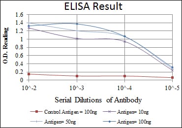
Red: Control Antigen (100ng); Purple: Antigen (10ng); Green: Antigen (50ng); Blue: Antigen (100ng);
For Research Use Only. Not for use in diagnostic procedures.