

PRKACG Primary Antibody
Item Information
Catalog #
Size
Price
Description
Cyclic AMP-dependent protein kinase (PKA) consists of two catalytic subunits and a regulatory subunit dimer. This gene encodes the gamma form of its catalytic subunit. The gene is intronless and is thought to be a retrotransposon derived from the gene for the alpha form of the PKA catalytic subunit.
Product Overview
Entrez GenelD
5568
Aliases
KAPG; PKACg
Clone#
2E4
Host / Isotype
Mouse / IgG1
Species Reactivity
Human
Immunogen
Purified recombinant fragment of human PRKACG expressed in E. Coli.
Formulation
Ascitic fluid containing 0.03% sodium azide.
Storage
Store at 4°C short term. Aliquot and store at -20°C long term. Avoid freeze/thaw cycles.
Product Applications
WB (Western Blot)
1/500 - 1/2000
IHC_P(Immunohistochemistry)
1/200 - 1/1000
FCM (Flow Cytometry)
1/200 - 1/400
ELISA
1/10000
References
1. Mol Cells. 2009 Jul 31;28(1):67-71.
2. J Clin Endocrinol Metab. 2009 Jul;94(7):2406-13.
2. J Clin Endocrinol Metab. 2009 Jul;94(7):2406-13.
Product Image
Western Blot

Figure 1: Western blot analysis using PRKACG mAb against human PRKACG (AA: 164-351) recombinant protein. (Expected MW is 47.1 kDa)
Immunohistochemical analysis

Figure 2: Immunohistochemical analysis of paraffin-embedded liver cancer tissues using PRKACG mouse mAb with DAB staining.
Immunohistochemical analysis

Figure 3: Immunohistochemical analysis of paraffin-embedded rectum cancer tissues using PRKACG mouse mAb with DAB staining.
Flow cytometric
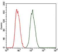
Figure 4: Flow cytometric analysis of MCF-7 cells using PRKACG mouse mAb (green) and negative control (red).
Elisa

Black line: Control Antigen (100 ng); Purple line: Antigen(10ng); Blue line: Antigen (50 ng); Red line: Antigen (100 ng);
For Research Use Only. Not for use in diagnostic procedures.